Explore
 Validate
 Learn
Learn Western blot
 ELISA
ELISAAntibody data
- Antibody Data
- Antigen structure
- References [0]
- Comments [0]
- Validations
- Western blot [2]
- Immunocytochemistry [1]
Submit
Validation data
Reference
Comment
Report error
- Product number
- MA1-34079 - Provider product page

- Provider
- Invitrogen Antibodies
- Product name
- PABP Monoclonal Antibody (10E10)
- Antibody type
- Monoclonal
- Antigen
- Recombinant full-length protein
- Description
- MA1-34079 detects PABP from human, chicken, rabbit and Xenopus laevis samples. Does not react wtih Drosophilia melanogaster or mouse samples.
- Reactivity
- Human, Chicken/Avian, Rabbit, Xenopus
- Host
- Mouse
- Isotype
- IgG
- Antibody clone number
- 10E10
- Vial size
- 50 µL
- Concentration
- 1.0 mg/mL
- Storage
- Store at 4°C short term. For long term storage, store at -20°C, avoiding freeze/thaw cycles.
No comments: Submit comment
Supportive validation
- Submitted by
- Invitrogen Antibodies (provider)
- Main image
- Experimental details
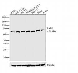
- Western blot analysis was performed on whole cell extracts (30 µg lysate) of A549 (Lane 1), HCT 116 (Lane 2), MCF7 (Lane 3), NTERA-2 cl.D1 (Lane 4), Hep G2 (Lane 5), HeLa (Lane 6) and A-431 (Lane 7). The blot was probed with Anti-PABP Monoclonal Antibody (Product # MA1-34079, 1:2000 dilution) and detected by chemiluminescence using Goat anti-Mouse IgG (H+L) Superclonal™ Secondary Antibody, HRP conjugate (Product # A28177, 0.25 µg/ml, 1:4000 dilution). A 70 kDa band corresponding to PABP was observed across the cell lines tested.

- Submitted by
- Invitrogen Antibodies (provider)
- Main image
- Experimental details
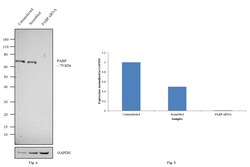
- Knockdown of PABP was achieved by transfecting HCT116 cells with PABP specific siRNAs (Silencer® select Product # s25666, s25664). Western blot analysis (Fig. a) was performed using whole cell extracts from the PABP knockdown cells (lane 3), non-specific scrambled siRNA transfected cells (lane 2) and untransfected cells (lane 1). The blot was probed with PABP Monoclonal Antibody (10E10) (Product # MA1-34079, 1:2000 dilution) and Goat anti-Mouse IgG (H+L) Superclonal™ Secondary Antibody, HRP conjugate (Product # A28177, 0.25µg/ml, 1:4000 dilution). Densitometric analysis of this western blot is shown in histogram (Fig. b). Decrease in signal upon siRNA mediated knock down confirms that antibody is specific to PABP.

Supportive validation
- Submitted by
- Invitrogen Antibodies (provider)
- Main image
- Experimental details
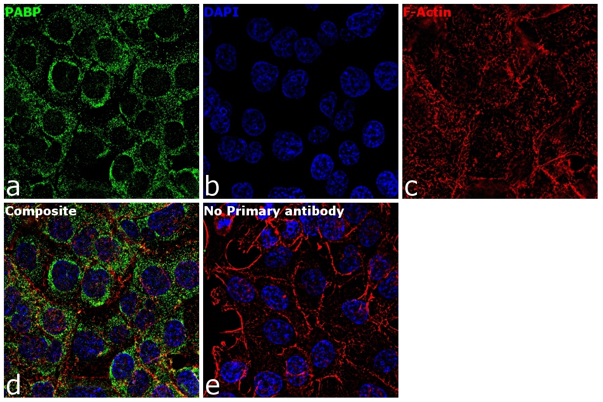
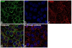
- Immunofluorescence analysis of PABP was performed using 70% confluent log phase HCT 116 cells. The cells were fixed with 4% paraformaldehyde for 10 minutes, permeabilized with 0.1% Triton™ X-100 for 15 minutes, and blocked with 1% BSA for 1 hour at room temperature. The cells were labeled with PABP Mouse Monoclonal Antibody(10E10) (Product # MA1-34079) at 5 µg/mL in 0.1% BSA, incubated at 4 degree Celsius overnight and then labeled with Goat anti-Mouse IgG (H+L) Superclonal™ Secondary Antibody, Alexa Fluor® 488 conjugate (Product # A28175) at a dilution of 1:2000 for 45 minutes at room temperature (Panel a: green). Nuclei (Panel b: blue) were stained with ProLong™ Diamond Antifade Mountant with DAPI (Product # P36962). F-actin (Panel c: red) was stained with Rhodamine Phalloidin (Product # R415, 1:300). Panel d represents the merged image showing cytoplasmic localization. Panel e represents control cells with no primary antibody to assess background. The images were captured at 60X magnification.