 Explore
 Validate
 Learn
Learn Western blot
Antibody data
- Antibody Data
- Antigen structure
- References [3]
- Comments [0]
- Validations
- Western blot [1]
- Immunocytochemistry [3]
- Immunohistochemistry [2]
- Other assay [3]
Submit
Validation data
Reference
Comment
Report error
- Product number
- MA1-079 - Provider product page

- Provider
- Invitrogen Antibodies
- Product name
- EIF2S1 Monoclonal Antibody (5A5)
- Antibody type
- Monoclonal
- Antigen
- Other
- Description
- MA1-079 has been successfully used in Western blot, immunohistochemistry, immunocytochemisty and immunofluorescent applications and reacts with human, monkey, and rat samples.
- Reactivity
- Human, Rat
- Host
- Mouse
- Isotype
- IgG
- Antibody clone number
- 5A5
- Vial size
- 100 µg
- Concentration
- 1 mg/mL
- Storage
- -20°C
Submitted references Dexmedetomidine Preconditioning Reduces Myocardial Ischemia-Reperfusion Injury in Rats by Inhibiting the PERK Pathway.
Fatal familial insomnia: mitochondrial and protein synthesis machinery decline in the mediodorsal thalamus.
The High-Risk Human Papillomavirus E6 Oncogene Exacerbates the Negative Effect of Tryptophan Starvation on the Development of Chlamydia trachomatis.
Chen Y, Cao S, Chen H, Yin C, Xu X, Yang Z
Arquivos brasileiros de cardiologia 2021 Dec;117(6):1134-1144
Arquivos brasileiros de cardiologia 2021 Dec;117(6):1134-1144
Fatal familial insomnia: mitochondrial and protein synthesis machinery decline in the mediodorsal thalamus.
Frau-Méndez MA, Fernández-Vega I, Ansoleaga B, Blanco Tech R, Carmona Tech M, Antonio Del Rio J, Zerr I, Llorens F, José Zarranz J, Ferrer I
Brain pathology (Zurich, Switzerland) 2017 Jan;27(1):95-106
Brain pathology (Zurich, Switzerland) 2017 Jan;27(1):95-106
The High-Risk Human Papillomavirus E6 Oncogene Exacerbates the Negative Effect of Tryptophan Starvation on the Development of Chlamydia trachomatis.
Sherchand SP, Ibana JA, Zea AH, Quayle AJ, Aiyar A
PloS one 2016;11(9):e0163174
PloS one 2016;11(9):e0163174
No comments: Submit comment
Supportive validation
- Submitted by
- Invitrogen Antibodies (provider)
- Main image
- Experimental details
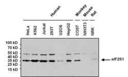
- Western blot analysis of eIF2S1 was performed by loading 80 µg of the indicated cell lysates onto a 4-20% Tris-HCl polyacrylamide gel. Proteins were transferred to a PVDF membrane and blocked with 5% BSA/TBST for at least 1 hour. The membrane was probed with an eIF2S1 monoclonal antibody (Product # MA1-079) at a dilution of 1:1000 overnight at 4°C on a rocking platform, washed in TBS-0.1%Tween 20, and probed with a goat anti-mouse IgG-HRP secondary antibody (Product # 31430) at a dilution of 1:20,000 for at least 1 hour. Chemiluminescent detection was performed using SuperSignal West Pico (Product # 34080).

Supportive validation
- Submitted by
- Invitrogen Antibodies (provider)
- Main image
- Experimental details
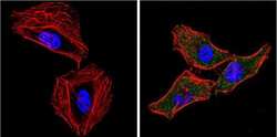
- Immunofluorescent analysis of eIF2S1 (green) showing positive staining in the cytoplasm of Hela cells (right) compared with a negative control in the absence of primary antibody (left). Formalin-fixed cells were permeabilized with 0.1% Triton X-100 in TBS for 5-10 minutes, blocked with 3% BSA-PBS for 30 minutes at room temperature and probed with an eIF2S1 monoclonal antibody (Product # MA1-079) in 3% BSA-PBS at a dilution of 1:100 and incubated overnight at 4 °C in a humidified chamber. Cells were washed with PBST and incubated with a DyLight 488-conjugated goat-anti-mouse IgG (H+L) secondary antibody in PBS at room temperature in the dark. F-actin (red) was stained with a fluorescent red phalloidin and nuclei (blue) were stained with DAPI for 5-10 minutes in the dark. Images were taken at a magnification of 60x.

- Submitted by
- Invitrogen Antibodies (provider)
- Main image

- Experimental details
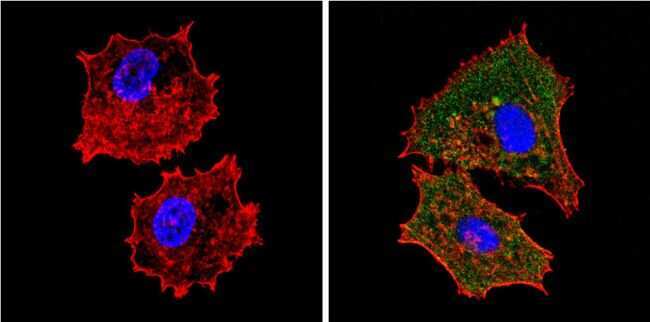
- Immunofluorescent analysis of eIF2S1 (green) showing positive staining in the cytoplasm of MCF-7 cells (right) compared with a negative control in the absence of primary antibody (left). Formalin-fixed cells were permeabilized with 0.1% Triton X-100 in TBS for 5-10 minutes, blocked with 3% BSA-PBS for 30 minutes at room temperature and probed with an eIF2S1 monoclonal antibody (Product # MA1-079) in 3% BSA-PBS at a dilution of 1:20 and incubated overnight at 4 °C in a humidified chamber. Cells were washed with PBST and incubated with a DyLight 488-conjugated goat-anti-mouse IgG (H+L) secondary antibody in PBS at room temperature in the dark. F-actin (red) was stained with a fluorescent red phalloidin and nuclei (blue) were stained with DAPI for 5-10 minutes in the dark. Images were taken at a magnification of 60x.
- Submitted by
- Invitrogen Antibodies (provider)
- Main image
- Experimental details
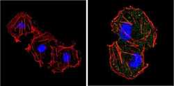
- Immunofluorescent analysis of eIF2S1 (green) showing positive staining in the cytoplasm of COS7 cells (right) compared with a negative control in the absence of primary antibody (left). Formalin-fixed cells were permeabilized with 0.1% Triton X-100 in TBS for 5-10 minutes, blocked with 3% BSA-PBS for 30 minutes at room temperature and probed with an eIF2S1 monoclonal antibody (Product # MA1-079) in 3% BSA-PBS at a dilution of 1:100 and incubated overnight at 4 °C in a humidified chamber. Cells were washed with PBST and incubated with a DyLight 488-conjugated goat-anti-mouse IgG (H+L) secondary antibody in PBS at room temperature in the dark. F-actin (red) was stained with a fluorescent red phalloidin and nuclei (blue) were stained with DAPI for 5-10 minutes in the dark. Images were taken at a magnification of 60x.

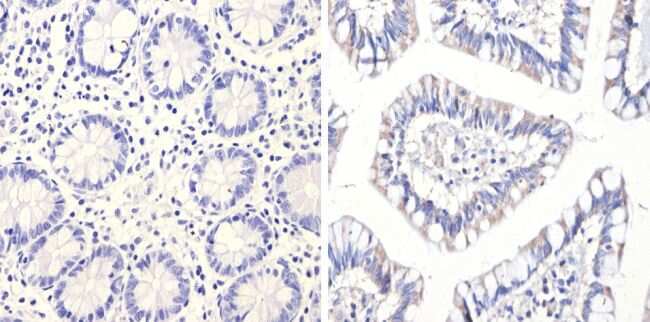
Supportive validation
- Submitted by
- Invitrogen Antibodies (provider)
- Main image
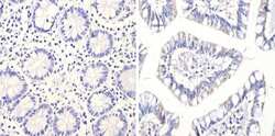
- Experimental details
- Immunohistochemistry analysis of eIF2S1 showing positive staining in the cytoplasm of paraffin-treated Human colon tissue (right) compared with a negative control in the absence of primary antibody (left). To expose target proteins, antigen retrieval method was performed using 10mM sodium citrate (pH 6.0), microwaved for 8-15 min. Following antigen retrieval, tissues were blocked in 3% H2O2-methanol for 15 min at room temperature, washed with ddH2O and PBS, and then probed with an eIF2S1 monoclonal antibody (Product # MA1-079) diluted by 3% BSA-PBS at a dilution of 1:20 overnight at 4°C in a humidified chamber. Tissues were washed extensively PBST and detection was performed using an HRP-conjugated secondary antibody followed by colorimetric detection using a DAB kit. Tissues were counterstained with hematoxylin and dehydrated with ethanol and xylene to prep for mounting.
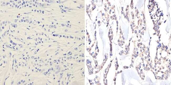
- Submitted by
- Invitrogen Antibodies (provider)
- Main image
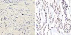
- Experimental details
- Immunohistochemistry analysis of eIF2S1 showing positive staining in the cytoplasm of paraffin-treated Human breast carcinoma (right) compared with a negative control in the absence of primary antibody (left). To expose target proteins, antigen retrieval method was performed using 10mM sodium citrate (pH 6.0), microwaved for 8-15 min. Following antigen retrieval, tissues were blocked in 3% H2O2-methanol for 15 min at room temperature, washed with ddH2O and PBS, and then probed with an eIF2S1 monoclonal antibody (Product # MA1-079) diluted by 3% BSA-PBS at a dilution of 1:20 overnight at 4°C in a humidified chamber. Tissues were washed extensively PBST and detection was performed using an HRP-conjugated secondary antibody followed by colorimetric detection using a DAB kit. Tissues were counterstained with hematoxylin and dehydrated with ethanol and xylene to prep for mounting.
Supportive validation
- Submitted by
- Invitrogen Antibodies (provider)
- Main image
- Experimental details
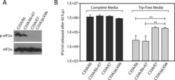
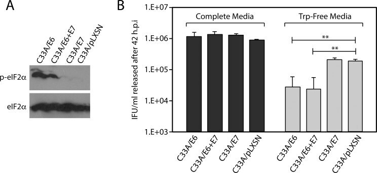
- Fig 6 Expression of the HPV16 E6 oncogene in C33A cells accentuates the effect of tryptophan starvation on C . trachomatis development. Stable C33A-derivative cell-lines were constructed by transducing C33A cells with a control retroviral vector (pLXSN), or pLXSN derivatives expressing the HPV16 E6, E6 & E7, or E7 oncogenes. Stable cell-lines were selected using G418 as described in the material and method section. A) Cell-lines were grown in tryptophan-free media for 12 hours after which immunoblots were used to query the phosphorylation status of eIF2alpha. Total eIF2alpha was used as a loading control. B) IFU/mL recovered at 42 h.p.i. after infected cells were grown in complete media or tryptophan free-media, as evaluated by infection of HeLa cells. As anticipated, growth in tryptophan-free media reduced IFU/mL recovered from all four cell-lines. IFU/mL recovery from C33A/E6 and C33A/E6+E7 cell-lines during tryptophan starvation were significantly lower than the IFU/mL recovered from the control C33A/pLXSN cell-line. The data represent three independent experiments (** indicates P < 0.01 by the Wilcoxon rank sum test).
- Submitted by
- Invitrogen Antibodies (provider)
- Main image
- Experimental details
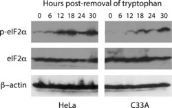
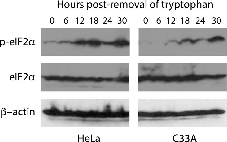
- Fig 2 Temporal evaluation of eIF2alpha phosphorylation in HeLa and C33A grown in tryptophan-free media. HeLa and C33A cells were plated in complete media, which was replaced with tryptophan-free media 24 hours post-plating. Cells were harvested every 6 hours and used to make extracts that were evaluated by immunoblot using antibodies against eIF2alpha or eIF2alpha phosphorylated on serine 51 (p-eIF2alpha). Immunoblots against beta-actin were performed as a loading control. Similar results were obtained from three independent experiments.
- Submitted by
- Invitrogen Antibodies (provider)
- Main image
- Experimental details
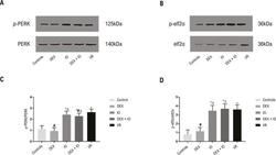
- Figura 6 - Efeito da DEX na expressao de p-PERK e p-eIF2alpha. (A, B) Western-blot detectou a expressao das proteinas PERK, p-PERK, eIF2alpha, p-eIF2alpha. (C, D) Analise da expressao de PERK, p-PERK, eIF2alpha, p-eIF2alpha no controle e no coracao isolado com I/R induzida. Dados sao apresentados como +- desvio padrao. n=6. * P
