 Explore
 Validate
 Learn
Learn Flow cytometry
Antibody data
- Antibody Data
- Antigen structure
- References [5]
- Comments [0]
- Validations
- Flow cytometry [1]
Submit
Validation data
Reference
Comment
Report error
- Product number
- MAB3478 - Provider product page

- Provider
- R&D Systems
- Product name
- Human CCR10 Antibody
- Antibody type
- Monoclonal
- Description
- Protein A or G purified from hybridoma culture supernatant. Detect human CCR10. Stains human CCR10-transfected cells but not irrelevant transfectants.
- Reactivity
- Human
- Host
- Rat
- Conjugate
- Unconjugated
- Antigen sequence
P46092- Isotype
- IgG
- Antibody clone number
- 314305
- Vial size
- 100 ug
- Concentration
- LYOPH
- Storage
- Use a manual defrost freezer and avoid repeated freeze-thaw cycles. 12 months from date of receipt, -20 to -70 °C as supplied. 1 month, 2 to 8 °C under sterile conditions after reconstitution. 6 months, -20 to -70 °C under sterile conditions after reconstitution.
Submitted references Skin Metabolites Define a New Paradigm in the Localization of Skin Tropic Memory T Cells.
The MEC1 and MEC2 lines represent two CLL subclones in different stages of progression towards prolymphocytic leukemia.
Microbe-specific unconventional T cells induce human neutrophil differentiation into antigen cross-presenting cells.
IL-1β promotes the differentiation of polyfunctional human CCR6+CXCR3+ Th1/17 cells that are specific for pathogenic and commensal microbes.
Vitamins A and D are potent inhibitors of cutaneous lymphocyte-associated antigen expression.
McCully ML, Collins PJ, Hughes TR, Thomas CP, Billen J, O'Donnell VB, Moser B
Journal of immunology (Baltimore, Md. : 1950) 2015 Jul 1;195(1):96-104
Journal of immunology (Baltimore, Md. : 1950) 2015 Jul 1;195(1):96-104
The MEC1 and MEC2 lines represent two CLL subclones in different stages of progression towards prolymphocytic leukemia.
Rasul E, Salamon D, Nagy N, Leveau B, Banati F, Szenthe K, Koroknai A, Minarovits J, Klein G, Klein E
PloS one 2014;9(8):e106008
PloS one 2014;9(8):e106008
Microbe-specific unconventional T cells induce human neutrophil differentiation into antigen cross-presenting cells.
Davey MS, Morgan MP, Liuzzi AR, Tyler CJ, Khan MWA, Szakmany T, Hall JE, Moser B, Eberl M
Journal of immunology (Baltimore, Md. : 1950) 2014 Oct 1;193(7):3704-3716
Journal of immunology (Baltimore, Md. : 1950) 2014 Oct 1;193(7):3704-3716
IL-1β promotes the differentiation of polyfunctional human CCR6+CXCR3+ Th1/17 cells that are specific for pathogenic and commensal microbes.
Duhen T, Campbell DJ
Journal of immunology (Baltimore, Md. : 1950) 2014 Jul 1;193(1):120-9
Journal of immunology (Baltimore, Md. : 1950) 2014 Jul 1;193(1):120-9
Vitamins A and D are potent inhibitors of cutaneous lymphocyte-associated antigen expression.
Yamanaka K, Dimitroff CJ, Fuhlbrigge RC, Kakeda M, Kurokawa I, Mizutani H, Kupper TS
The Journal of allergy and clinical immunology 2008 Jan;121(1):148-157.e3
The Journal of allergy and clinical immunology 2008 Jan;121(1):148-157.e3
No comments: Submit comment
Supportive validation
- Submitted by
- R&D Systems (provider)
- Main image
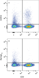
- Experimental details
- Detection of CCR10 in Human Blood Lymphocytes by Flow Cytometry. Human peripheral blood lymphocytes were stained with Mouse Anti-Human CD3 epsilon APC-conjugated Monoclonal Antibody (Catalog # FAB100A) and either (A) Rat Anti-Human CCR10 Monoclonal Antibody (Catalog # MAB3478) or (B) Rat IgG2A Isotype Control (Catalog # MAB006) followed by Phycoerythrin-conjugated Anti-Rat IgG Secondary Antibody (Catalog # F0105B). View our protocol for Staining Membrane-associated Proteins.
