Explore
 Validate
 Learn
Learn Western blot
Antibody data
- Antibody Data
- Antigen structure
- References [2]
- Comments [0]
- Validations
- Western blot [2]
- Immunocytochemistry [3]
- Chromatin Immunoprecipitation [2]
- Other assay [1]
Submit
Validation data
Reference
Comment
Report error
- Product number
- MA5-23510 - Provider product page

- Provider
- Invitrogen Antibodies
- Product name
- Phospho-RNA pol II CTD (Ser2) Monoclonal Antibody
- Antibody type
- Monoclonal
- Antigen
- Other
- Reactivity
- Human
- Host
- Mouse
- Isotype
- IgG
- Vial size
- 50 µg
- Concentration
- 1 mg/mL
- Storage
- -20°C or -80°C if preferred
Submitted references Epigenetic Suppression of HIV in Myeloid Cells by the BRD4-Selective Small Molecule Modulator ZL0580.
Structure-guided drug design identifies a BRD4-selective small molecule that suppresses HIV.
Alamer E, Zhong C, Liu Z, Niu Q, Long F, Guo L, Gelman BB, Soong L, Zhou J, Hu H
Journal of virology 2020 May 18;94(11)
Journal of virology 2020 May 18;94(11)
Structure-guided drug design identifies a BRD4-selective small molecule that suppresses HIV.
Niu Q, Liu Z, Alamer E, Fan X, Chen H, Endsley J, Gelman BB, Tian B, Kim JH, Michael NL, Robb ML, Ananworanich J, Zhou J, Hu H
The Journal of clinical investigation 2019 Jul 22;129(8):3361-3373
The Journal of clinical investigation 2019 Jul 22;129(8):3361-3373
No comments: Submit comment
Supportive validation
- Submitted by
- Invitrogen Antibodies (provider)
- Main image
- Experimental details
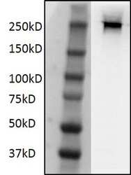
- Western blot analysis of POLR2A was performed by loading 50 µg of J-lat full (10.6) whole cell lysate in reducing sample buffer onto a 4-15% tris-glycine gel. Proteins were transferred to PVDF and then blocked in blocking buffer (TBST+5% non-fat milk) for one hour at room temperature. POLR2A was detected using an anti-POLR2A monoclonal antibody (Product # MA5-23510) at a dilution of 1:1,000 overnight at 4°C on a rocking platform. Goat anti Mouse HRP-conjugated secondary antibody was diluted at 1:2000 in 1xTBST buffer with 5% non fat milk and incubated for 1 hour at room temperature. Data courtesy of Antibody Data Exchange Program.

- Submitted by
- Invitrogen Antibodies (provider)
- Main image
- Experimental details
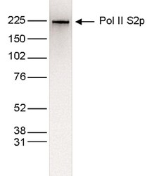
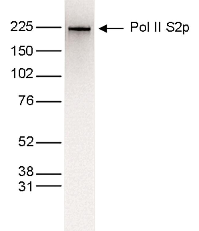
- Nuclear extracts (25 µg) from HeLa cells were analyzed by Western Blot using a RNA Polymerase II S2p monoclonal antibody (Product # MA5-23510) at a dilution of 1:1,000 in TBS-Tween containing 5% skimmed milk. The position of the protein of interest is indicated on the right; the marker (kDa) is shown on the left.
Supportive validation
- Submitted by
- Invitrogen Antibodies (provider)
- Main image

- Experimental details
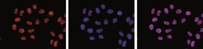
- Immunofluorescent detection of RNA Polymerase II S2p in HeLa cells stained with RNA Polymerase II S2p monoclonal antibody (Product # MA5-23510). Cells were fixed with methanol and blocked with PBS/Triton X-100 containing 5% normal goat serum and 1% BSA. The cells were immunofluorescently labelled with the Pol II S2p antibody (left) at a dilution of 1:500 in blocking solution followed by an anti-mouse antibody conjugated to AlexaFluor 594. The middle panel shows staining of the nuclei with DAPI. A merge of the two stainings is shown on the right.
- Submitted by
- Invitrogen Antibodies (provider)
- Main image

- Experimental details
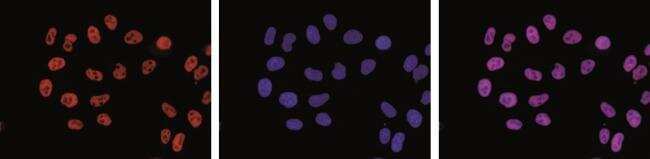
- Immunofluorescence analysis of Phospho-RNA pol II CTD (Ser2) Monoclonal Antibody was performed using 70% confluent log phase HCT 116 cells. The cells were fixed with 4% paraformaldehyde for 10 minutes, permeabilized with 0.1% Triton™ X-100 for 15 minutes, and blocked with 2% BSA for 45 minutes at room temperature. The cells were labeled with Phospho-RNA pol II CTD (Ser2) Monoclonal Antibody (Product # MA5-23510) at 1:100 dilution in 0.1% BSA, incubated at 4 degree celsius overnight and then labeled with Donkey anti-Mouse IgG (H+L) Highly Cross-Adsorbed Secondary Antibody, Alexa Fluor Plus 488 (Product # A32766), (1:2000 dilution), for 45 minutes at room temperature (Panel a: Green). Nuclei (Panel b:Blue) were stained with ProLong™ Diamond Antifade Mountant with DAPI (Product # P36962). F-actin (Panel c: Red) was stained with Rhodamine Phalloidin (Product # R415, 1:300 dilution). Panel d represents the merged image showing Nuclear localization. Panel e represents control cells with no primary antibody to assess background. The images were captured at 60X magnification.

- Submitted by
- Invitrogen Antibodies (provider)
- Main image

- Experimental details
- Immunofluorescent detection of RNA Polymerase II S2p in HeLa cells stained with RNA Polymerase II S2p monoclonal antibody (Product # MA5-23510). Cells were fixed with methanol and blocked with PBS/Triton X-100 containing 5% normal goat serum and 1% BSA. The cells were immunofluorescently labelled with the Pol II S2p antibody (left) at a dilution of 1:500 in blocking solution followed by an anti-mouse antibody conjugated to AlexaFluor 594. The middle panel shows staining of the nuclei with DAPI. A merge of the two stainings is shown on the right.
Supportive validation
- Submitted by
- Invitrogen Antibodies (provider)
- Main image

- Experimental details
- ChIP assays were performed on human HeLa cells using a RNA Polymerase II S2p monoclonal antibody (Product # MA5-23510) and optimized PCR primer pairs for qPCR. ChIP was performed using sheared chromatin from 1 million cells. A titration consisting of 1, 2, 5 and 10 µg of antibody per ChIP experiment was analyzed. IgG (2 µg/IP) was used as a negative IP control. Quantitative PCR was performed with primers specific for the coding region of the constitutively expressed GAPDH and ACTB genes, used as positive controls, and for exon 2 of the inactive myoglobin (MB) gene and the Sat2 satellite repeat, used as negative controls. Figure 1 shows the recovery, expressed as a % of input (the relative amount of immunoprecipitated DNA compared to input DNA after qPCR analysis).

- Submitted by
- Invitrogen Antibodies (provider)
- Main image
- Experimental details
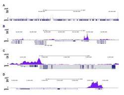
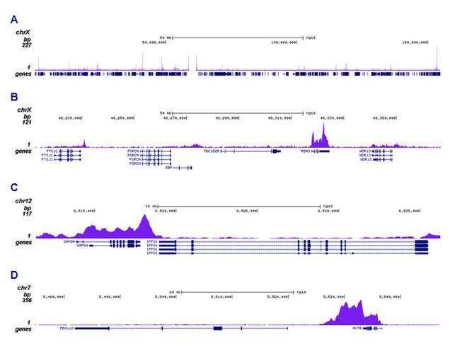
- ChIP was performed on sheared chromatin from 1 million HeLaS3 cells using 1 µg RNA Polymerase II S2p monoclonal antibody (Product # MA5-23510). The IP'd DNA was subsequently analyzed on a Genome Analyzer. The 36 bp tags were aligned to the human genome using the ELAND algorithm. Figure 2 shows the peak distribution along the complete sequence and a 150 kb region of the X-chromosome (figure 2A and B, respectively), and in a two genomic regions surrounding the GAPDH and ACTB positive control genes (figure 2C and D).
Supportive validation
- Submitted by
- Invitrogen Antibodies (provider)
- Main image
- Experimental details
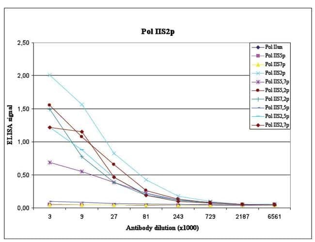
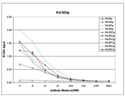
- To test the specificity, an ELISA was performed using a serial dilution of RNA Polymerase II S2p monoclonal antibody (Product # MA5-23510). The wells were coated with peptides containing the unmodified C-terminal repeat sequence as well as different phosphorylated peptides. Figure 3 shows the specificity of the antibody for the S2 phosphorylation.