Explore
 Validate
 Learn
Learn Western blot
Antibody data
- Antibody Data
- Antigen structure
- References [0]
- Comments [0]
- Validations
- Western blot [3]
- Immunocytochemistry [1]
- Immunohistochemistry [3]
Submit
Validation data
Reference
Comment
Report error
- Product number
- PA5-28448 - Provider product page

- Provider
- Invitrogen Antibodies
- Product name
- HP1 beta Polyclonal Antibody
- Antibody type
- Polyclonal
- Antigen
- Synthetic peptide
- Description
- Recommended positive controls: Raji.
- Concentration
- 1 mg/mL
No comments: Submit comment
Supportive validation
- Submitted by
- Invitrogen Antibodies (provider)
- Main image
- Experimental details
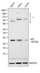
- Western blot was performed using Anti- HP1 Polyclonal Antibody (Product # PA5-28448) and a 25 kDa band corresponding to HP1 was observed across the cell lines along with additional bands between 60-160kDa. Modified whole cell extracts (30ug) of MCF7 (Lane 1), HeLa (Lane 2) and A-431 (Lane 3) were electrophoresed using Novex® NuPAGE® 4-12 % Bis-Tris gel (Product # NP0322BOX). Resolved proteins were then transferred onto a nitrocellulose membrane (Product # IB23001) by iBlot® 2 Dry Blotting System (Product # IB21001). The blot was probed with the primary antibody (1:1000 dilution) and detected by chemiluminescence with Goat anti-Rabbit IgG (H+L), Superclonal™ Recombinant Secondary Antibody, HRP (Product # A27036, 1:4000 dilution) using the iBright FL 1000 (Product # A32752). Chemiluminescent detection was performed using Novex® ECL Chemiluminescent Substrate Reagent Kit (Product # WP20005)..

- Submitted by
- Invitrogen Antibodies (provider)
- Main image
- Experimental details
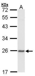
- Western Blot using HP1 beta Polyclonal Antibody (Product # PA5-28448). Sample (30 µg of whole cell lysate). Lane A: Raji. 12% SDS PAGE. HP1 beta Polyclonal Antibody (Product # PA5-28448) diluted at 1:1,000.

- Submitted by
- Invitrogen Antibodies (provider)
- Main image

- Experimental details
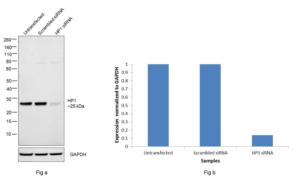
- Knockdown of HP1 was achieved by transfecting MCF7 with HP1 specific siRNAs (Silencer® select Product # s21549). Western blot analysis (Fig. a) was performed using whole cell extracts from the HP1 knockdown cells (lane 3), non-specific scrambled siRNA transfected cells (lane 2) and untransfected cells (lane 1). The blot was probed with HP1 Monoclonal Antibody (Product # PA5-28448, 2µg/ml) and Goat anti-Mouse IgG (H+L) Superclonal™ Recombinant Secondary Antibody, HRP (Product # A28177, 1:4000 dilution). Densitometric analysis of this western blot is shown in histogram (Fig. b). Decrease in signal upon siRNA mediated knock down confirms that antibody is specific to HP1.
Supportive validation
- Submitted by
- Invitrogen Antibodies (provider)
- Main image
- Experimental details
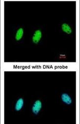
- Immunofluorescent analysis of CBX1/HP1 beta in paraformaldehyde-fixed HeLa cells using a CBX1/HP1 beta polyclonal antibody (Product # PA5-28448) at a 1:200 dilution.

Supportive validation
- Submitted by
- Invitrogen Antibodies (provider)
- Main image
- Experimental details
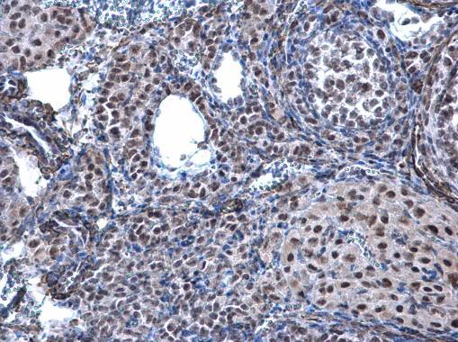
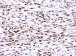
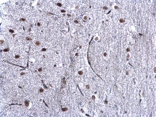
- CBX1/HP1 beta antibody detects CBX1 protein at nucleus on C2C12 xenograft by immunohistochemical analysis. Sample: Paraffin-embedded C2C12 xenograft. CBX1/HP1 beta antibody (Product # PA5-28448) dilution: 1:500. Antigen Retrieval: EDTA based buffer, pH 8.0, 15 min.
- Submitted by
- Invitrogen Antibodies (provider)
- Main image
- Experimental details
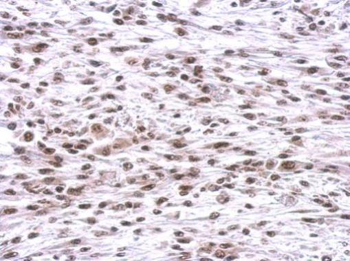
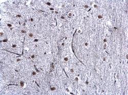
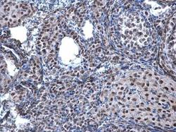
- CBX1/HP1 beta antibody detects CBX1/HP1 beta protein at nucleus on mouse ovary by immunohistochemical analysis. Sample: Paraffin-embedded mouse ovary. CBX1/HP1 beta antibody (Product # PA5-28448) diluted at 1:500. Antigen Retrieval: EDTA based buffer, pH 8.0, 15 min.
- Submitted by
- Invitrogen Antibodies (provider)
- Main image
- Experimental details
- CBX1/HP1 beta antibody detects CBX1/HP1 beta protein at nucleus on mouse ovary by immunohistochemical analysis. Sample: Paraffin-embedded mouse ovary. CBX1/HP1 beta antibody (Product # PA5-28448) diluted at 1:500. Antigen Retrieval: EDTA based buffer, pH 8.0, 15 min.