Explore
 Validate
 Learn
Learn Western blot
Antibody data
- Antibody Data
- Antigen structure
- References [0]
- Comments [0]
- Validations
- Western blot [3]
- Immunocytochemistry [2]
- Immunohistochemistry [3]
Submit
Validation data
Reference
Comment
Report error
- Product number
- PA5-40134 - Provider product page

- Provider
- Invitrogen Antibodies
- Product name
- Glutaminase C (GAC) Polyclonal Antibody
- Antibody type
- Polyclonal
- Antigen
- Recombinant full-length protein
- Description
- This antibody specifically recognizes GAC but not the KGA isoform.
- Concentration
- 0.5 mg/mL
No comments: Submit comment
Supportive validation
- Submitted by
- Invitrogen Antibodies (provider)
- Main image
- Experimental details
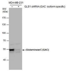
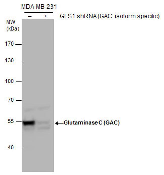
- Western blot analysis and shRNA knock-down of Glutaminase C in non-transfected (-) and GLS1 (GAC isoform specific) shRNA-transfected MDA-MB-231 whole cell extracts (30 µg). Samples were separated by 7.5% SDS-PAGE and the membrane was probed with a Glutaminase C polyclonal antibody (Product # PA5-40134) at a dilution of 1:5000.
- Submitted by
- Invitrogen Antibodies (provider)
- Main image
- Experimental details
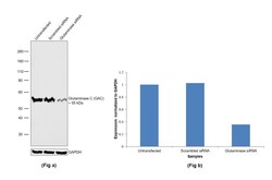
- Knockdown of Glutaminase was achieved by transfecting ACHN with Glutaminase specific siRNAs (Silencer® select Product # s5840, s5839). Western blot analysis (Fig. a) was performed using whole cell extracts from the Glutaminase knockdown cells (lane 3), non-specific scrambled siRNA transfected cells (lane 2) and untransfected cells (lane 1). The blot was probed with Glutaminase C (GAC) Polyclonal Antibody (Product # PA5-40134, 1:5000 dilution) and Goat anti-Rabbit IgG (H+L) Superclonal™ Recombinant Secondary Antibody, HRP (Product # A27036, 0.25 µg/mL, 1:4000 dilution). Densitometric analysis of this western blot is shown in histogram (Fig. b). Decrease in signal upon siRNA mediated knock down confirms that antibody is specific to Glutaminase.

- Submitted by
- Invitrogen Antibodies (provider)
- Main image
- Experimental details
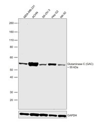
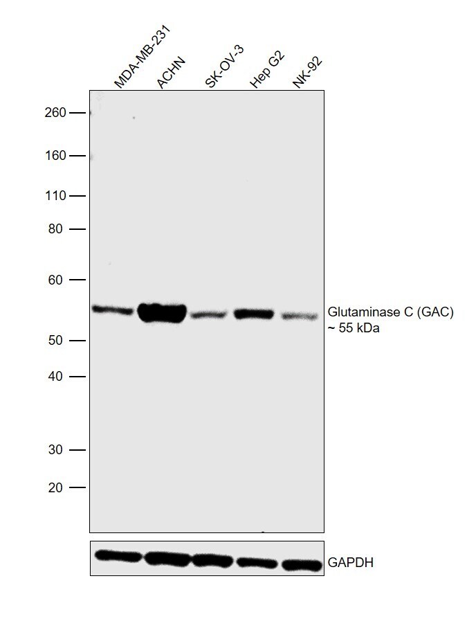
- Western blot was performed using Anti-Glutaminase C (GAC) Polyclonal Antibody (Product # PA5-40134) and a 55 kDa band corresponding to Glutaminase was observed across all the cell lines tested. Whole cell extracts (30 µg lysate) of MDA-MB-231 (Lane 1), ACHN (Lane 2), SK-OV-3 (Lane 3), Hep G2 (Lane 4) and NK-92 (Lane 5) were electrophoresed using NuPAGE™ 4-12% Bis-Tris Protein Gel (Product # NP0322BOX). Resolved proteins were then transferred onto a nitrocellulose membrane (Product # IB23001) by iBlot® 2 Dry Blotting System (Product # IB21001). The blot was probed with the primary antibody (1:5000 dilution) and detected by chemiluminescence with Goat anti-Rabbit IgG (H+L) Superclonal™ Recombinant Secondary Antibody, HRP (Product # A27036, 1:4000 dilution) using the iBright FL 1000 (Product # A32752). Chemiluminescent detection was performed using Novex® ECL Chemiluminescent Substrate Reagent Kit (Product # WP20005).
Supportive validation
- Submitted by
- Invitrogen Antibodies (provider)
- Main image

- Experimental details
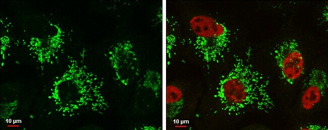
- Glutaminase C antibody detects Glutaminase C protein at mitochondria by immunofluorescent analysis. Sample: A549 cells were fixed in 4% paraformaldehyde at RT for 15 min. Green: Glutaminase C protein stained by Glutaminase C antibody (Product # PA5-40134) diluted at 1:500. Red: Histone H3K9ac (acetyl Lys9), a nucleus marker, stained by Histone H3K9ac (acetyl Lys9) antibody [GT464] (Product # MA5-31512) diluted at 1:500. Blue: Hoechst 33342 staining. Scale bar = 10 μm.
- Submitted by
- Invitrogen Antibodies (provider)
- Main image
- Experimental details
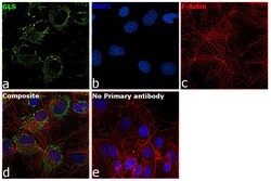
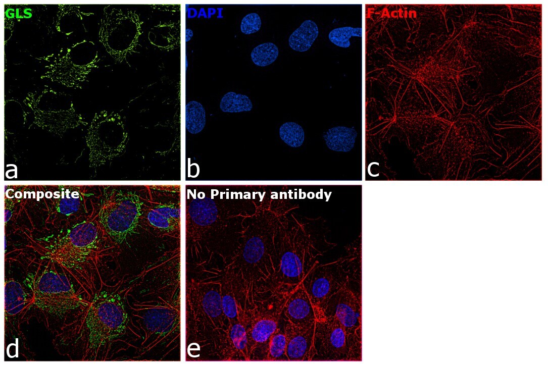
- Immunofluorescence analysis of GLS was performed using 70% confluent log phase Hep G2 cells. The cells were fixed with 4% paraformaldehyde for 10 minutes, permeabilized with 0.1% Triton™ X-100 for 15 minutes, and blocked with 2% BSA for 1 hour at room temperature. The cells were labeled with Glutaminase C (GAC) Polyclonal Antibody (Product # PA5-40134) at 1:100 dilution in 0.1% BSA, incubated at 4 degree Celsius overnight and then with Donkey anti-Rabbit IgG (H+L) Highly Cross-Adsorbed Secondary Antibody, Alexa Fluor Plus 488 (Product # A32790) at a dilution of 1:2000 for 45 minutes at room temperature (Panel a: green). Nuclei (Panel b: blue) were stained with SlowFade® Gold Antifade Mountant with DAPI (Product # S36938). F-actin (Panel c: red) was stained with Rhodamine Phalloidin (Product # R415, 1:300). Panel d represents the merged image showing mitochondrial pattern in cytoplasm. Panel e represents control cells with no primary antibody to assess background. The images were captured at 60X magnification.
Supportive validation
- Submitted by
- Invitrogen Antibodies (provider)
- Main image
- Experimental details
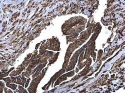
- Glutaminase C antibody detects Glutaminase C protein at cytoplasm on human breast carcinoma by immunohistochemical analysis. Sample: Paraffin-embedded human breast carcinoma. Glutaminase C antibody (Product # PA5-40134) diluted at 1:500. Antigen Retrieval: Citrate buffer, pH 6.0, 15 min.
- Submitted by
- Invitrogen Antibodies (provider)
- Main image
- Experimental details
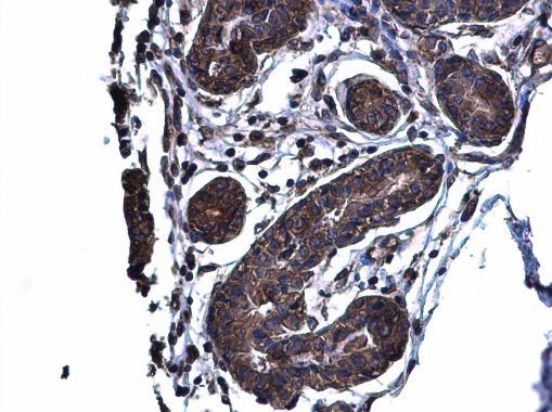
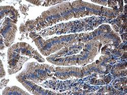
- Glutaminase C antibody detects Glutaminase C protein at cytoplasm on human colon carcinoma by immunohistochemical analysis. Sample: Paraffin-embedded human colon carcinoma. Glutaminase C antibody (Product # PA5-40134) diluted at 1:500. Antigen Retrieval: Citrate buffer, pH 6.0, 15 min.

- Submitted by
- Invitrogen Antibodies (provider)
- Main image
- Experimental details
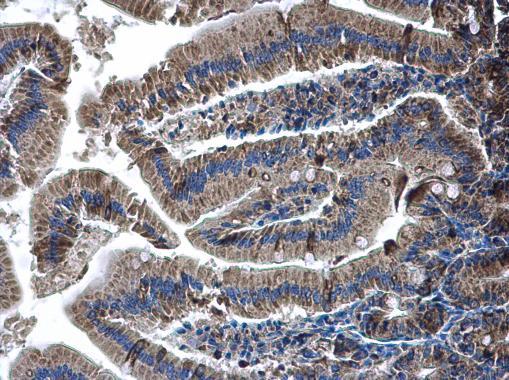
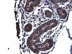
- Glutaminase C antibody detects Glutaminase C protein at cytoplasm on mouse duidenum by immunohistochemical analysis. Sample: Paraffin-embedded mouse duidenum. Glutaminase C antibody (Product # PA5-40134) diluted at 1:500. Antigen Retrieval: EDTA based buffer, pH 8.0, 15 min.