 Explore
 Validate
 Learn
Learn Western blot
Antibody data
- Antibody Data
- Antigen structure
- References [1]
- Comments [0]
- Validations
- Western blot [2]
- Immunocytochemistry [1]
- Other assay [1]
Submit
Validation data
Reference
Comment
Report error
- Product number
- 710449 - Provider product page

- Provider
- Invitrogen Antibodies
- Product name
- SRC Recombinant Polyclonal Antibody (14HCLC)
- Antibody type
- Polyclonal
- Antigen
- Synthetic peptide
- Description
- Recombinant rabbit polyclonal antibodies are unique offerings from Thermo Fisher Scientific. They are comprised of a selection of multiple different recombinant monoclonal antibodies, providing the best of both worlds - the sensitivity of polyclonal antibodies with the specificity of monoclonal antibodies - all delivered with the consistency only found in a recombinant antibody. While functionally the same as a polyclonal antibody - recognizing multiple epitope sites on the target and producing higher detection sensitivity for low abundance targets - a recombinant rabbit polyclonal antibody has a known mixture of light and heavy chains. The exact population can be produced in every lot, circumventing the biological variability typically associated with polyclonal antibody production.
- Reactivity
- Human, Mouse
- Host
- Rabbit
- Isotype
- IgG
- Antibody clone number
- 14HCLC
- Vial size
- 100 µg
- Concentration
- 0.5 mg/mL
- Storage
- Store at 4°C short term. For long term storage, store at -20°C, avoiding freeze/thaw cycles.
Submitted references Microenvironmental Factors Drive Tenascin C and Src Cooperation to Promote Invadopodia Formation in Ewing Sarcoma.
Hawkins AG, Julian CM, Konzen S, Treichel S, Lawlor ER, Bailey KM
Neoplasia (New York, N.Y.) 2019 Oct;21(10):1063-1072
Neoplasia (New York, N.Y.) 2019 Oct;21(10):1063-1072
No comments: Submit comment
Supportive validation
- Submitted by
- Invitrogen Antibodies (provider)
- Main image

- Experimental details
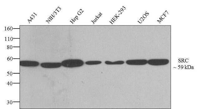
- Western blot analysis of SRC was performed by loading 20 µg of A431 (lane1), NIH/3T3 (lane2), Hep G2 (lane3), Jurkat (lane4), HEK-293 (lane5), U2OS (lane6) and MCF7 (lane7) cell lysates using Novex®NuPAGE®4-12% Bis-Tris gel (Product # NP0321BOX), XCell SureLock Electrophoresis System (Product # EI0002), Novex® Sharp Pre-Stained Protein Standard (Product # LC5800), and iBlot® Dry Blotting System (Product # IB21001). Proteins were transferred to a nitrocellulose membrane and blocked with 5% skim milk for 1 hour at room temperature. SRC was detected at ~59 kDa using SRC Recombinant Rabbit Polyclonal Antibody (Product # 710449) at 1-2 µg/mL in 2.5% skim milk at 4°C overnight on a rocking platform. Goat anti-Rabbit IgG - HRP Secondary Antibody (Product # G-21234) at 1:5000 dilution was used and chemiluminescent detection was performed using Pierce™ ECL Western blotting Substrate (Product # 32106).
- Submitted by
- Invitrogen Antibodies (provider)
- Main image
- Experimental details
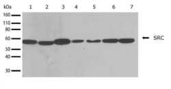
- Western blot analysis of SRC in whole cell extracts from A431, NIH-3T3, HepG2, Jurkat, HEK293, U20S, and MCF-7 (lanes 1-7 respectively) using a SRC Recombinant Rabbit Polyclonal Antibody (Product # 710449) at a dilution of 1 µg/mL. Detection was performed using an HRP-conjugated Goat anti-Rabbit secondary antibody followed by chemiluminescence (ECL). Results show a band at ~59kDa.

Supportive validation
- Submitted by
- Invitrogen Antibodies (provider)
- Main image
- Experimental details
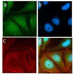
- Immunofluorescent analysis of SRC in HeLa cells using a SRC Recombinant Rabbit Polyclonal Antibody (Product # 710449) followed by detection using an Alexa Fluor 488-conjugated Goat anti-Rabbit secondary antibody (green) (Image A). Nuclei were stained using DAPI (Image B) and actin stained with Alexa Fluor 594 phalloidin (red) (image C). Image D is a composite image showing cytoplasmic and nuclear localization of SRC.

Supportive validation
- Submitted by
- Invitrogen Antibodies (provider)
- Main image
- Experimental details
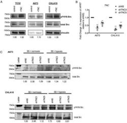
- Figure 2 Cell intrinsic and extrinsic tenascin C enhances Src phosphorylation in Ewing sarcoma. A, TC32, A673 and CHLA10 Ewing sarcoma cells were treated with 3 micro-Molar recombinant tenascin C (TNC) or vehicle control for 24 hours. Lysates were subjected to western blot analysis for p-Y416 Src, total Src and vinculin. B, A673 and CHLA10 cells were stably transduced with lentivirus containing non-silencing hairpin (shNS) or two different TNC hairpins (shTNC3 and shTNC5). Knockdown was confirmed via qRT-PCR for TNC . C, p-Src and Src expression were measured via western blot in both A673 (top panels) and CHLA10 (bottom panels) cells transduced with control (shNS) or TNC knockdown (shTNC3 and shTNC5) and in the presence of serum starvation (SS), normoxia or hypoxia. Figure 2
