Explore
 Validate
 Learn
Learn Western blot
Antibody data
- Antibody Data
- Antigen structure
- References [2]
- Comments [0]
- Validations
- Western blot [4]
- Immunocytochemistry [4]
- Immunohistochemistry [1]
Submit
Validation data
Reference
Comment
Report error
- Product number
- MA5-15140 - Provider product page

- Provider
- Invitrogen Antibodies
- Product name
- Phospho-S6 (Ser235, Ser236) Monoclonal Antibody (G.22.6)
- Antibody type
- Monoclonal
- Antigen
- Synthetic peptide
- Description
- It is not recommended to aliquot this antibody.
- Reactivity
- Human, Mouse, Rat
- Host
- Rabbit
- Isotype
- IgG
- Antibody clone number
- G.22.6
- Vial size
- 100 µL
- Concentration
- 72 µg/mL
- Storage
- -20°C
Submitted references Essential role of glucokinase in the protection of pancreatic β cells to the glucose energetic status.
Autophagy plays a protective role in endoplasmic reticulum stress-mediated pancreatic β cell death.
Marqués P, Kamitz A, Bartolomé A, Burillo J, Martínez H, Jiménez B, Fernández-Rhodes M, Guillén C, Benito M
Cell death discovery 2019;5:138
Cell death discovery 2019;5:138
Autophagy plays a protective role in endoplasmic reticulum stress-mediated pancreatic β cell death.
Bartolome A, Guillen C, Benito M
Autophagy 2012 Dec;8(12):1757-68
Autophagy 2012 Dec;8(12):1757-68
No comments: Submit comment
Supportive validation
- Submitted by
- Invitrogen Antibodies (provider)
- Main image

- Experimental details
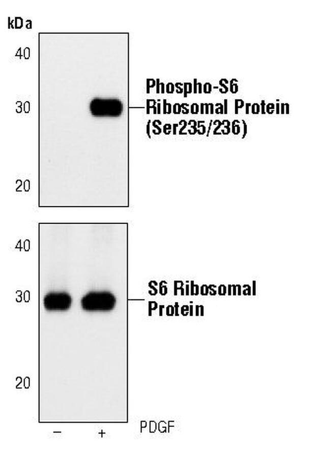
- Western blot analysis of Phospho-S6 Ribosomal Protein pSer235/236 in extracts from NIH/3T3 cells, untreated or PDGF-treated (100 ng/mL, 20 min), using Phospho-S6 Ribosomal Protein pSer235/236 monoclonal antibody (Product # MA5-15140) (upper) or a S6 Ribosomal Protein monoclonal antibody (lower).

- Submitted by
- Invitrogen Antibodies (provider)
- Main image

- Experimental details
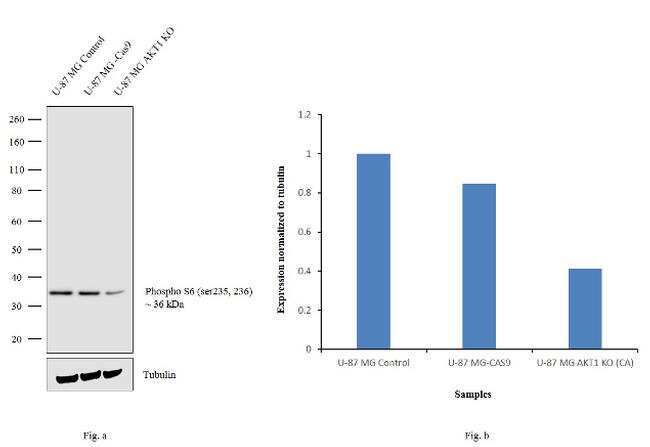
- Western blot analysis of Phospho-S6 kinase(Ser235, Ser236) (Fig. a) was performed by loading 20 µg of U-87 MG Control (lane 1), U-87 MG Cas9 (lane 2), U-87 MG AKT1 KO (lane 3) membrane extracts. Phospho-S6 kinase (Ser235, Ser236) was detected at 36 kDa using Phospho-S6 kinase(Ser235, Ser236) monoclonal Antibody (G.22.6) (Product # MA5-15140, 1:1000 dilution) and Goat anti-Mouse IgG (H+L) Superclonal™ Secondary Antibody HRP conjugate (Product # A28177, 1:4000 dilution). Densitometric analysis of this western blot is shown in histogram (Fig. b). Loss of signal in CRISPR mediated knockout (KO) confirms that antibody is specific to Phospho-S6 kinase(Ser235, Ser236).
- Submitted by
- Invitrogen Antibodies (provider)
- Main image

- Experimental details
- Western blot analysis was performed on whole cell extracts (30 µg lysate) of U-2 OS (Lane 1), U-2 OS treated with IGF (50 ng/mL for 6 hr) (Lane 2) and U-2 OS treated with Rapamycin (50nM for 4 hr) followed by treatment with IGF (50 ng/mL for 6hr) (Lane 3). The blot was probed with anti-Phospho-S6 (Ser235, Ser236) Monoclonal Antibody (Product # MA5-15140, 1:500 dilution) and detected by chemiluminescence using Goat anti-Rabbit IgG (H+L) Superclonal™ Secondary Antibody, HRP conjugate (Product # A27036, 0.25 µg/mL, 1:4000 dilution). A 32 kDa band corresponding to Phospho-S6 (Ser235, Ser236) was enhanced upon IGF treatment and was decreased upon rapamycin treatment on the cell line tested.

- Submitted by
- Invitrogen Antibodies (provider)
- Main image

- Experimental details
- Western blot analysis of Phospho-S6 Ribosomal Protein pSer235/236 in extracts from NIH/3T3 cells, untreated or PDGF-treated (100 ng/mL, 20 min), using Phospho-S6 Ribosomal Protein pSer235/236 monoclonal antibody (Product # MA5-15140) (upper) or a S6 Ribosomal Protein monoclonal antibody (lower).
Supportive validation
- Submitted by
- Invitrogen Antibodies (provider)
- Main image

- Experimental details
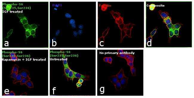
- Immunofluorescence analysis of Phospho-S6 (Ser235,Ser236) was performed using 70% confluent log phase U-2 OS cells treated with 50 ng/mL IGF-1 for 6hrs. The cells were fixed with 4% paraformaldehyde for 10 minutes, permeabilized with 0.1% Triton™ X-100 for 10 minutes, and blocked with 1% BSA for 1 hour at room temperature. The cells were labeled with Phospho-S6 (Ser235,Ser236) Monoclonal Antibody (Product # MA5-15140) at 1:100 dilution in 0.1% BSA, incubated overnight at 4 degree Celsius and then labeled with Goat anti-Rabbit IgG (H+L) Superclonal™ Secondary Antibody, Alexa Fluor® 488 conjugate (Product # A27034) at a dilution of 1:2000 for 45 minutes at room temperature (Panel a: green). Nuclei (Panel b: blue) were stained with SlowFade® Gold Antifade Mountant with DAPI (Product # S36938). F-actin (Panel c: red) was stained with Rhodamine Phalloidin (Product # R415, 1:300). Panel d represents the merged image showing cytoplasmic localization. Panel e represents cells treated with antagonist, Rapamycin (50 nM for 4hrs) followed by IGF (50 ng/mL for 6hrs), showing reduced expression of phospho-S6. Panel f shows the untreated cells showing basal expression of phospho S6. Panel g shows control cells with no primary antibody to assess background. The images were captured at 60X magnification.
- Submitted by
- Invitrogen Antibodies (provider)
- Main image
- Experimental details
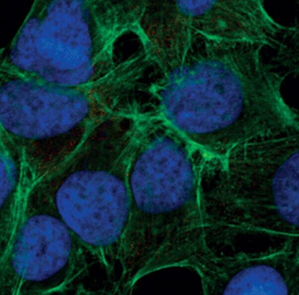
- Immunofluorescent analysis of Phospho-S6 Ribosomal Protein pSer235/236 in HeLa cells, serum-starved for 20 hrs, using a Phospho-S6 Ribosomal Protein pSer235/236 monoclonal antibody (Product # MA5-15140) (red). Actin filaments have been labeled with FITC-conjugated phalloidin. DNA is labeled using a fluorescent blue dye.

- Submitted by
- Invitrogen Antibodies (provider)
- Main image

- Experimental details
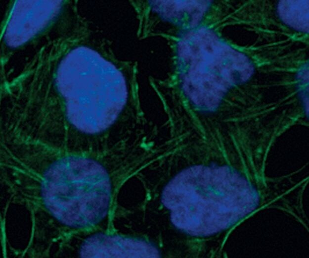
- Immunofluorescent analysis of Phospho-S6 Ribosomal Protein pSer235/236 in HeLa cells, 20% serum-treated after preincubation with Rapamycin (FRAP/mTOR Inhibitor), using a Phospho-S6 Ribosomal Protein pSer235/236 monoclonal antibody (Product # MA5-15140) (red). Actin filaments have been labeled with FITC-conjugated phalloidin. DNA is labeled using a fluorescent blue dye.
- Submitted by
- Invitrogen Antibodies (provider)
- Main image

- Experimental details
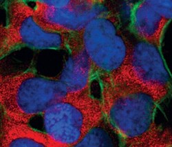
- Immunofluorescent analysis of Phospho-S6 Ribosomal Protein pSer235/236 in HeLa cells, 20% serum-treated, using a Phospho-S6 Ribosomal Protein pSer235/236 monoclonal antibody (Product # MA5-15140) (red). Actin filaments have been labeled with FITC-conjugated phalloidin. DNA is labeled using a fluorescent blue dye.
Supportive validation
- Submitted by
- Invitrogen Antibodies (provider)
- Main image

- Experimental details
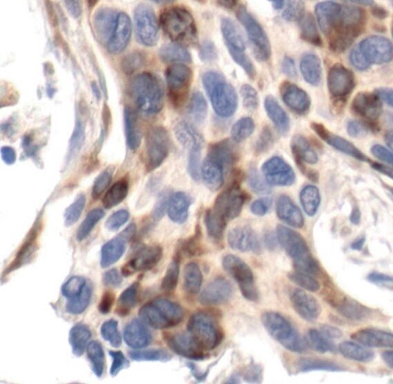
- Immunohistochemical analysis of Phospho-S6 Ribosomal Protein pSer235/236 in paraffin-embedded human breast carcinoma using a Phospho-S6 Ribosomal Protein pSer235/236 monoclonal antibody (Product # MA5-15140).