Explore
 Validate
 Learn
Learn Western blot
Antibody data
- Antibody Data
- Antigen structure
- References [0]
- Comments [0]
- Validations
- Western blot [2]
- Immunocytochemistry [1]
Submit
Validation data
Reference
Comment
Report error
- Product number
- PA5-38116 - Provider product page

- Provider
- Invitrogen Antibodies
- Product name
- Anti-Phospho-HDAC5 (Ser259) Polyclonal Antibody
- Antibody type
- Polyclonal
- Antigen
- Synthetic peptide
- Reactivity
- Human, Mouse
- Host
- Rabbit
- Isotype
- IgG
- Vial size
- 100 µg
- Concentration
- 1 mg/mL
- Storage
- -20°C
No comments: Submit comment
Supportive validation
- Submitted by
- Invitrogen Antibodies (provider)
- Main image
- Experimental details
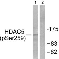
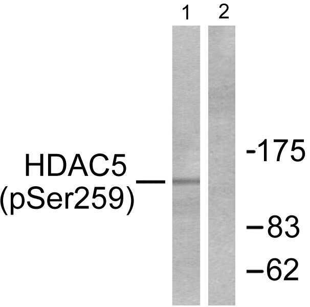
- Western blot analysis of Phospho-HDAC5 pSer259 in extracts from HepG2 cells using a Phospho-HDAC5 pSer259 polyclonal antibody (Product # PA5-38116).
- Submitted by
- Invitrogen Antibodies (provider)
- Main image
- Experimental details
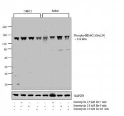
- Western blot analysis was performed on modified whole cell extracts (1% SDS) (30 µg lysate) of IMR32 (Lane 1), IMR32 treated with Ionomycin (0.5 uM for 1 min) (Lane 2), IMR32 treated with Ionomycin (0.5 uM for 5 min) (Lane 3), IMR32 treated with Ionomycin (0.5 uM for 60 min) (Lane 4), Jurkat (Lane 5), Jurkat treated with Ionomycin (0.5 uM for 1 min) (Lane 6), Jurkat treated with Ionomycin (0.5 uM for 5 min) (Lane 7), Jurkat treated with Ionomycin (0.5 uM for 60 min) (Lane 8). The blot was probed with Anti-Phospho-HDAC5 (Ser259) Polyclonal Antibody (Product # PA5-38116, 1:1000 dilution) and detected by chemiluminescence using Goat anti-Rabbit IgG (H+L) Superclonal™ Secondary Antibody, HRP conjugate (Product # A27036, 0.25 µg/ml, 1:4000 dilution). A 122 kDa band corresponding to Phospho-HDAC5 (Ser259) was observed across the cell lines tested and showed treatment dependent difference in RAW 264.7 cell line upon Ionomycin treatment with varying time points.

Supportive validation
- Submitted by
- Invitrogen Antibodies (provider)
- Main image
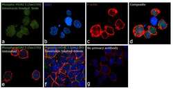
- Experimental details
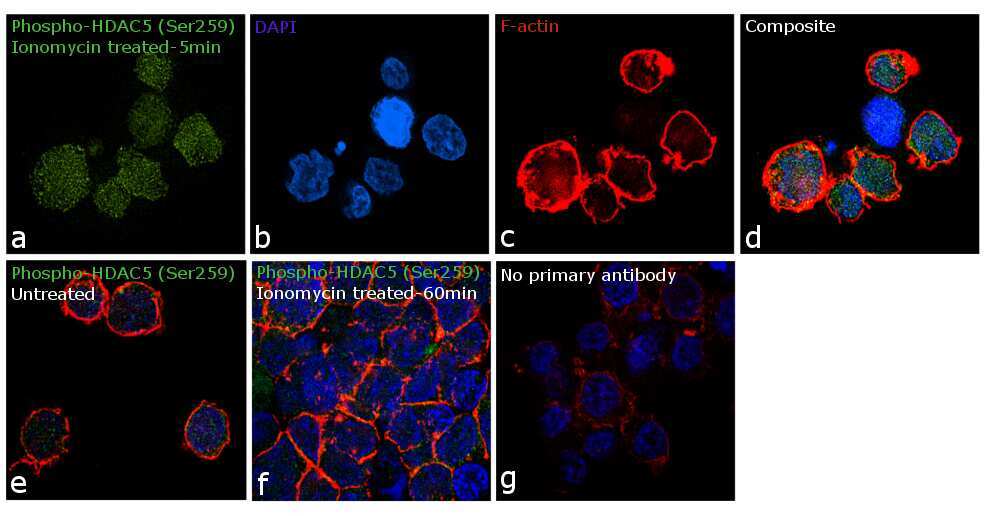
- Immunofluorescence analysis of Phospho-HDAC5 (Ser259) was performed using 70% confluent log phase Jurkat cells treated with Ionomycin. The cells were fixed with 4% paraformaldehyde for 10 minutes, permeabilized with 0.1% Triton™ X-100 for 15 minutes, and blocked with 1% BSA for 1 hour at room temperature. The cells were labeled with Phospho-HDAC5 (Ser259) Polyclonal Antibody(Product # PA5-38116) at 1:100 dilution in 0.1% BSA, incubated at 4 degree Celsius overnight and then labeled with Goat anti-Rabbit IgG (H+L) Superclonal™ Secondary Antibody, Alexa Fluor® 488 conjugate (Product # A27034) at a dilution of 1:2000 for 45 minutes at room temperature (Panel a: green). Nuclei (Panel b: blue) were stained with ProLong™ Diamond Antifade Mountant with DAPI (Product # P36962). F-actin (Panel c: red) was stained with Rhodamine Phalloidin (Product # R415, 1:300). Panel d represents the merged image showing increased expression upon Ionomycin treatment for 5min. Panel e represents the control cells showing weak nuclear staining. Panel f shows decreased staining upon Ionomycin treatment for 60 min. Panel g represents control cells with no primary antibody to assess background. The images were captured at 60X magnification.