Explore
 Validate
 Learn
Learn Western blot
 Immunohistochemistry
Antibody data
- Antibody Data
- Antigen structure
- References [2]
- Comments [0]
- Validations
- Immunohistochemistry [2]
Submit
Validation data
Reference
Comment
Report error
- Product number
- 36-0200 - Provider product page

- Provider
- Invitrogen Antibodies
- Product name
- Anti-VEGF Receptor 3 Polyclonal Antibody
- Antibody type
- Polyclonal
- Antigen
- Other
- Reactivity
- Human
- Host
- Rabbit
- Isotype
- IgG
- Vial size
- 100 µg
- Concentration
- 0.25 mg/ml
- Storage
- -20°C
Submitted references Expression of factors involved in the regulation of angiogenesis in the full-term human placenta: Effects of in vitro fertilization.
Immunohistochemical expression and distribution of VEGFR-3 in malignant mesothelioma.
Li C, Zhang Y, Tang L, Zhao H, Gao C, Gao L, Cui Y, Liu J
Reproductive biology 2016 Jun;16(2):104-12
Reproductive biology 2016 Jun;16(2):104-12
Immunohistochemical expression and distribution of VEGFR-3 in malignant mesothelioma.
Filho AL, Baltazar F, Bedrossian C, Michael C, Schmitt FC
Diagnostic cytopathology 2007 Dec;35(12):786-91
Diagnostic cytopathology 2007 Dec;35(12):786-91
No comments: Submit comment
Supportive validation
- Submitted by
- Invitrogen Antibodies (provider)
- Main image
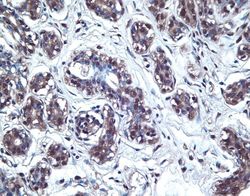
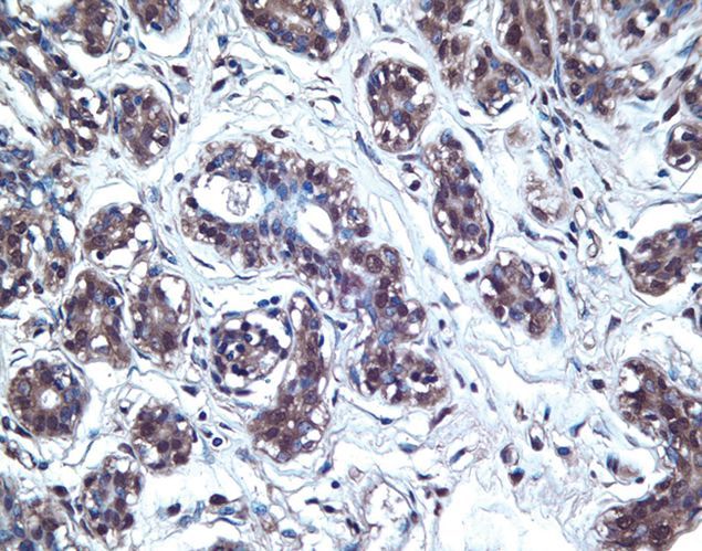
- Experimental details
- Immunohistochemical staining of breast carcinoma tissue using Rb anti-VEGF Receptor-3 (C-term) (PAD: ZMD.251) (Product # 36-0200).
- Submitted by
- Invitrogen Antibodies (provider)
- Main image
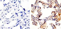
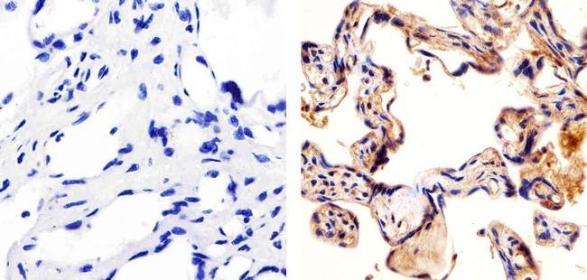
- Experimental details
- Immunohistochemistry analysis of FLT-4/VEGFR3 showing staining in the cytoplasm of paraffin-embedded human placenta tissue (right) compared to a negative control without primary antibody (left). To expose target proteins, antigen retrieval was performed using 10mM sodium citrate (pH 6.0), microwaved for 8-15 min. Following antigen retrieval, tissues were blocked in 3% H2O2-methanol for 15 min at room temperature, washed with ddH2O and PBS, and then probed with a Anti- FLT-4/VEGFR3 Polyclonal Antibody (Product # 36-0200) diluted in 3% BSA-PBS at a dilution of 1:20 overnight at 4°C in a humidified chamber. Tissues were washed extensively in PBST and detection was performed using an HRP-conjugated secondary antibody followed by colorimetric detection using a DAB kit. Tissues were counterstained with hematoxylin and dehydrated with ethanol and xylene to prep for mounting.