 Explore
 Validate
 Learn
Learn Western blot
Antibody data
- Antibody Data
- Antigen structure
- References [6]
- Comments [0]
- Validations
- Western blot [1]
- Immunocytochemistry [2]
Submit
Validation data
Reference
Comment
Report error
- Product number
- MA1-2001 - Provider product page

- Provider
- Invitrogen Antibodies
- Product name
- beta Catenin Monoclonal Antibody (9F2)
- Antibody type
- Monoclonal
- Antigen
- Purifed from natural sources
- Description
- MA1-2001 detects beta catenin from human samples.
- Antibody clone number
- 9F2
- Concentration
- 1 mg/mL
Submitted references microRNA-133b represses the progression of lung cancer through inhibiting SOX9/β-catenin signaling pathway.
Wnt Signaling Separates the Progenitor and Endocrine Compartments during Pancreas Development.
Interaction of alpha-actinin with the cadherin/catenin cell-cell adhesion complex via alpha-catenin.
Interaction of alpha-actinin with the cadherin/catenin cell-cell adhesion complex via alpha-catenin.
Identification of plakoglobin domains required for association with N-cadherin and alpha-catenin.
Identification of plakoglobin domains required for association with N-cadherin and alpha-catenin.
Liu S, Li S, Yu X, Wang Q, Sun H
International journal of clinical and experimental pathology 2020;13(9):2270-2279
International journal of clinical and experimental pathology 2020;13(9):2270-2279
Wnt Signaling Separates the Progenitor and Endocrine Compartments during Pancreas Development.
Sharon N, Vanderhooft J, Straubhaar J, Mueller J, Chawla R, Zhou Q, Engquist EN, Trapnell C, Gifford DK, Melton DA
Cell reports 2019 May 21;27(8):2281-2291.e5
Cell reports 2019 May 21;27(8):2281-2291.e5
Interaction of alpha-actinin with the cadherin/catenin cell-cell adhesion complex via alpha-catenin.
Knudsen KA, Soler AP, Johnson KR, Wheelock MJ
The Journal of cell biology 1995 Jul;130(1):67-77
The Journal of cell biology 1995 Jul;130(1):67-77
Interaction of alpha-actinin with the cadherin/catenin cell-cell adhesion complex via alpha-catenin.
Knudsen KA, Soler AP, Johnson KR, Wheelock MJ
The Journal of cell biology 1995 Jul;130(1):67-77
The Journal of cell biology 1995 Jul;130(1):67-77
Identification of plakoglobin domains required for association with N-cadherin and alpha-catenin.
Sacco PA, McGranahan TM, Wheelock MJ, Johnson KR
The Journal of biological chemistry 1995 Aug 25;270(34):20201-6
The Journal of biological chemistry 1995 Aug 25;270(34):20201-6
Identification of plakoglobin domains required for association with N-cadherin and alpha-catenin.
Sacco PA, McGranahan TM, Wheelock MJ, Johnson KR
The Journal of biological chemistry 1995 Aug 25;270(34):20201-6
The Journal of biological chemistry 1995 Aug 25;270(34):20201-6
No comments: Submit comment
Supportive validation
- Submitted by
- Invitrogen Antibodies (provider)
- Main image
- Experimental details
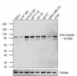
- Western blot analysis was performed on whole cell extracts (30 µg lysate) of HeLa (Lane 1), U-87 MG (Lane 2), COLO 205 (Lane 3), HCT 116 (lane 4), HT-29 (lane 5), MCF7 (lane 6), MDA-MB-231 (lane 7) and A549 (lane 8). The blots were probed with Anti-beta Catenin Mouse Monoclonal Antibody (Product # MA1-2001, 1-3 µg/mL) and detected by chemiluminescence using Goat anti-Mouse IgG (H+L) Secondary Antibody, HRP conjugate (Product # 62-6520, 1:4000 dilution). A 85 kDa band corresponding to beta Catenin was observed across cell lines tested. Known quantity of protein samples were electrophoresed using Novex® NuPAGE® 4-12 % Bis-Tris gel (Product # NP0322BOX), XCell SureLock™ Electrophoresis System (Product # EI0002) and Novex® Sharp Pre-Stained Protein Standard (Product # LC5800). Resolved proteins were then transferred onto a nitrocellulose membrane with iBlot® 2 Dry Blotting System (Product # IB21001). The membrane was probed with the relevant primary and secondary Antibody following blocking with 5 % skimmed milk. Chemiluminescent detection was performed using Pierce™ ECL Western Blotting Substrate (Product # 32106).

Supportive validation
- Submitted by
- Invitrogen Antibodies (provider)
- Main image
- Experimental details
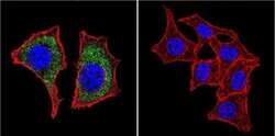
- Immunofluorescent analysis of Beta-Catenin using Beta-Catenin Monoclonal antibody (9F2) (Product # MA1-2001) shows staining in HeLa cells. Beta-Catenin staining (green), F-Actin staining with Phalloidin (red) and nuclei with DAPI (blue) is shown. Cells were grown on chamber slides and fixed with formaldehyde prior to staining. Cells were probed without (control) or with or an antibody recognizing Beta-Catenin (Product # MA1-2001) at a dilution of 1:20 over night at 4 °C, washed with PBS and incubated with a DyLight-488 conjugated secondary antibody (Product # 35552 for GAR, Product # 35503 for GAM). Images were taken at 60X magnification.
- Submitted by
- Invitrogen Antibodies (provider)
- Main image
- Experimental details
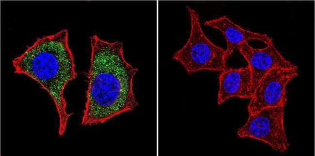
- Immunofluorescent analysis of Beta-Catenin using Beta-Catenin Monoclonal antibody (9F2) (Product # MA1-2001) shows staining in U251 glioma cells. Beta-Catenin staining (green), F-Actin staining with Phalloidin (red) and nuclei with DAPI (blue) is shown. Cells were grown on chamber slides and fixed with formaldehyde prior to staining. Cells were probed without (control) or with or an antibody recognizing Beta-Catenin (Product # MA1-2001) at a dilution of 1:20 over night at 4 °C, washed with PBS and incubated with a DyLight-488 conjugated secondary antibody (Product # 35552 for GAR, Product # 35503 for GAM). Images were taken at 60X magnification.
