 Explore
 Validate
 Learn
Learn Western blot
 Other assay
Antibody data
- Antibody Data
- Antigen structure
- References [2]
- Comments [0]
- Validations
- Other assay [2]
Submit
Validation data
Reference
Comment
Report error
- Product number
- MA1-35681 - Provider product page

- Provider
- Invitrogen Antibodies
- Product name
- MRP4 Monoclonal Antibody (M4I-10)
- Antibody type
- Monoclonal
- Antigen
- Other
- Reactivity
- Human
- Host
- Rat
- Isotype
- IgG
- Antibody clone number
- M4I-10
- Vial size
- 1 mL
- Concentration
- 0.1 mg/mL
- Storage
- 4° C
Submitted references Photodynamic Priming Improves the Anti-Migratory Activity of Prostaglandin E Receptor 4 Antagonist in Cancer Cells In Vitro.
Identification of MRP4/ABCC4 as a Target for Reducing the Proliferation of Pancreatic Ductal Adenocarcinoma Cells by Modulating the cAMP Efflux.
Sorrin AJ, Liu C, Cicalo J, Reader J, Najafali D, Zhang Y, Roque DM, Huang HC
Cancers 2021 Oct 20;13(21)
Cancers 2021 Oct 20;13(21)
Identification of MRP4/ABCC4 as a Target for Reducing the Proliferation of Pancreatic Ductal Adenocarcinoma Cells by Modulating the cAMP Efflux.
Carozzo A, Yaneff A, Gómez N, Di Siervi N, Sahores A, Diez F, Attorresi AI, Rodríguez-González Á, Monczor F, Fernández N, Abba M, Shayo C, Davio C
Molecular pharmacology 2019 Jul;96(1):13-25
Molecular pharmacology 2019 Jul;96(1):13-25
No comments: Submit comment
Supportive validation
- Submitted by
- Invitrogen Antibodies (provider)
- Main image
- Experimental details
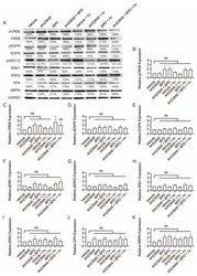
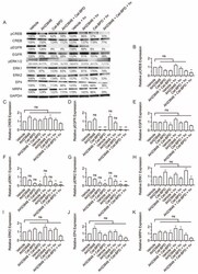
- Figure 2 Western blot analysis of p-CREB, CREB, p-EGFR, EGFR, p-ERK1/2, ERK1, ERK2, EP4, and MRP4 in OVCAR-5 cells. Cells were treated with the indicated agents for 24 h, then light-activated (0.1 J/cm 2 , 10 mW/cm 2 ) or maintained in dark conditions. After 24 h, cells were agonized with EGF (50 ng/mL) and PGE2 (1 uM) for 10 min, then whole extracts were collected and analyzed using Western blot. ( A ) Representative Western blot images and ( B - K ) relative densitometric bar graphs of phosphorylated and total proteins were shown. Results are normalized to the vehicle control group. Statistical analysis was performed using a one-way ANOVA and post hoc Tukey's test. Percentages below each band represent the average change in intensity relative to the vehicle control across all experiments. For pERK1 and pERK2 bands, the first number corresponds to pERK1, and the second number corresponds to pERK2. Error bars represent the standard error of the mean. * p
- Submitted by
- Invitrogen Antibodies (provider)
- Main image
- Experimental details
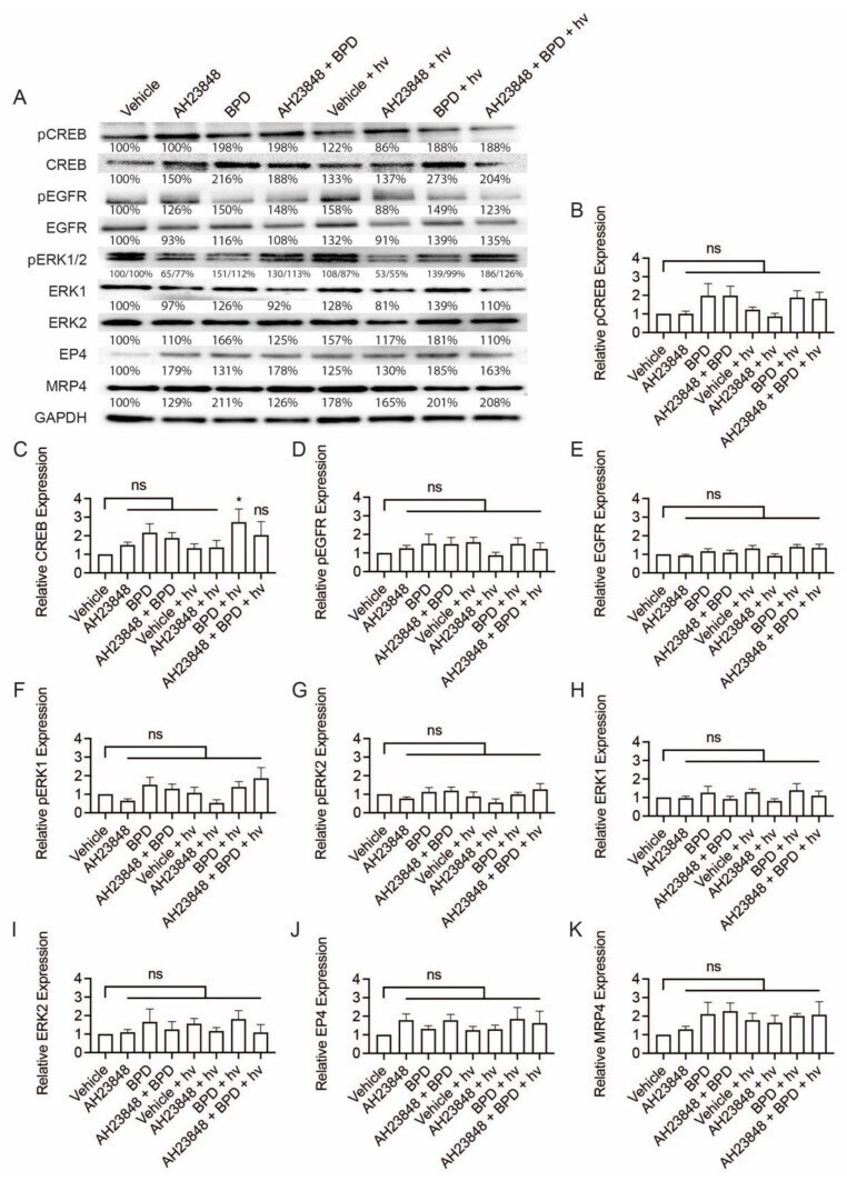
- Figure 5 Western blot analysis of p-CREB, CREB, p-EGFR, EGFR, p-ERK1/2, ERK1, ERK2, EP4, and MRP4 in OVCAR-5 cells. Cells were treated with the indicated agents for 24 h, then light-activated (0.1 J/cm 2 , 10 mW/cm 2 ) or maintained in dark conditions. After 24 h, cells were agonized with EGF (50 ng/mL) and PGE 2 (1 uM) for 10 min, then whole extracts were collected and analyzed using Western blot. ( A ) Representative Western blot images and ( B - K ) relative densitometric bar graphs of phosphorylated and total proteins were shown. Results are normalized to the vehicle control group. Statistical analysis was performed using a one-way ANOVA and post hoc Tukey's test. Percentages below each band represent the average change in intensity relative to the vehicle control across all experiments. For pERK1 and pERK2 bands, the first number corresponds to pERK1, and the second number corresponds to pERK2. Error bars represent the standard error of the mean. * p
