Explore
 Validate
 Learn
Learn Western blot
 Immunoprecipitation
Antibody data
- Antibody Data
- Antigen structure
- References [0]
- Comments [0]
- Validations
- Western blot [1]
- Immunocytochemistry [2]
Submit
Validation data
Reference
Comment
Report error
- Product number
- MA5-16703 - Provider product page

- Provider
- Invitrogen Antibodies
- Product name
- Huntingtin Monoclonal Antibody (HDB4E10)
- Antibody type
- Monoclonal
- Antigen
- Recombinant full-length protein
- Description
- Increased cytoplasmic staining, relative to nuclear, has been reported using formaldehyde as a fixative compared with acetone/methanol. A suggested positive control for immunohistochemical applications is brain.
- Reactivity
- Human, Mouse, Rabbit
- Host
- Mouse
- Isotype
- IgG
- Antibody clone number
- HDB4E10
- Vial size
- 100 µg
- Concentration
- 1 mg/mL
- Storage
- Store at 4°C short term. For long term storage, store at -20°C, avoiding freeze/thaw cycles.
No comments: Submit comment
Supportive validation
- Submitted by
- Invitrogen Antibodies (provider)
- Main image

- Experimental details
- Western blot analysis of Huntingtin in normal human cerebral cortex total protein extract. Samples were icubated in Huntingtin Monoclonal antibody (Product # MA5-16703). Run on a 3-12.5% gradient SDS-PAGE gel.

Supportive validation
- Submitted by
- Invitrogen Antibodies (provider)
- Main image
- Experimental details
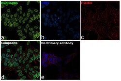
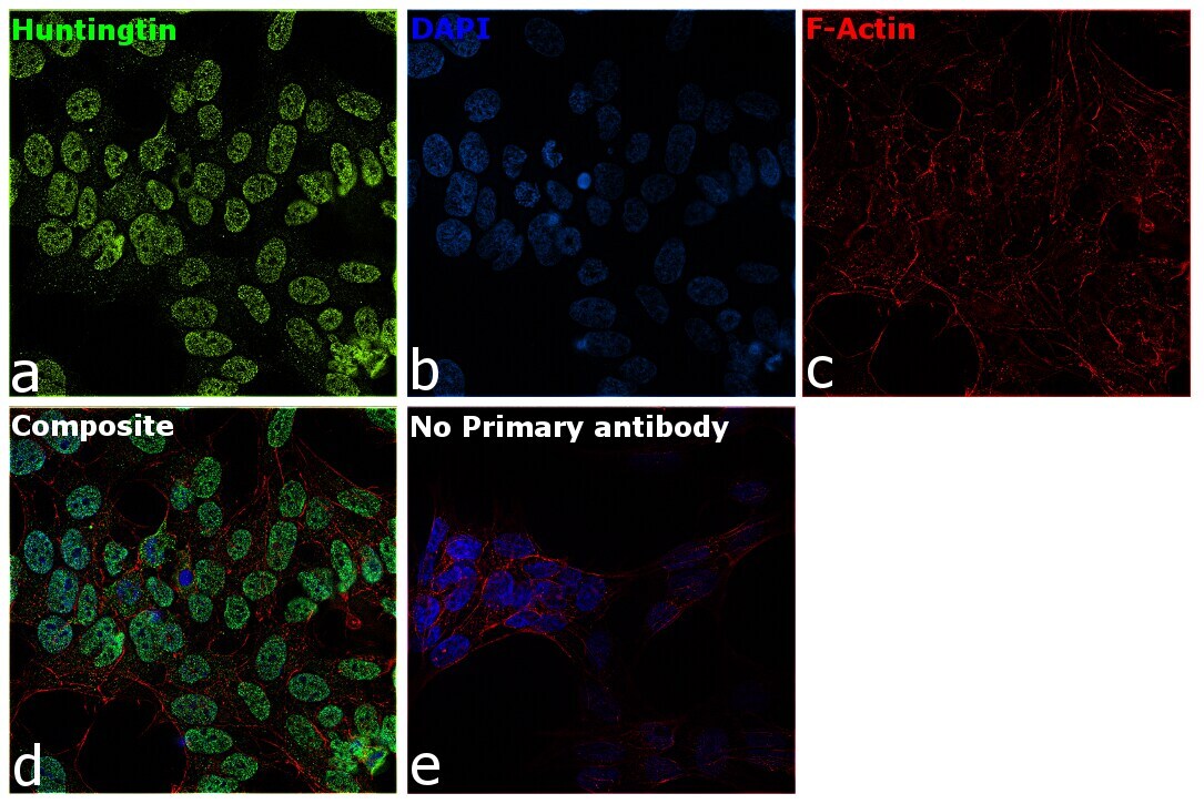
- Immunofluorescence analysis of Huntingtin was performed using 70% confluent log phase SH-SY5Y cells. The cells were fixed with 4% paraformaldehyde for 10 minutes, permeabilized with 0.1% Triton™ X-100 for 15 minutes, and blocked with 2% BSA for 1 hour at room temperature. The cells were labeled with Huntingtin Monoclonal Antibody (Product # MA5-16703) at 1:100 dilution in 0.1% BSA, incubated at 4 degree celsius overnight and then with Goat anti-Rabbit IgG (H+L), Superclonal™ Recombinant Secondary Antibody, Alexa Fluor 488 conjugate (Product # A27034) at a dilution of 1:2000 for 45 minutes at room temperature (Panel a: green). Nuclei (Panel b: blue) were stained with SlowFade® Gold Antifade Mountant with DAPI (Product # S36938). F-actin (Panel c: red) was stained with Rhodamine Phalloidin (Product # R415, 1:300). Panel d represents the merged image showing predominantly nuclear and faint cytosolic localization. Panel e represents no primary antibody to assess background. The images were captured at 60X magnification.
- Submitted by
- Invitrogen Antibodies (provider)
- Main image
- Experimental details
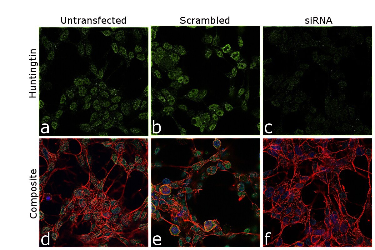
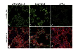
- Knockdown of HTT was achieved by transfecting SH-SY5Y cells with HTT specific siRNA (Silencer® select Product # s6492, s6491). Immunofluorescence analysis was performed on U-87 MG cells (untransfected, panel a,d), transfected with non-specific scrambled siRNA (panels b,e) and transfected with HTT specific siRNA (panel c,f). Cells were fixed, permeabilized, and labelled with Huntingtin Monoclonal Antibody (HDB4E10) (Product # MA5-16703, 1:100 dilution), followed by Goat anti-Mouse IgG (H+L) Superclonal™ Recombinant Secondary Antibody, Alexa Fluor® 488 (Product # A28175, 1:2000). Nuclei (blue) were stained using ProLong™ Diamond Antifade Mountant with DAPI (Product # P36962), and Rhodamine Phalloidin (Product # R415, 1:300) was used for cytoskeletal F-actin (red) staining. Loss of specific signal was observed upon siRNA mediated knockdown (panel c,f) confirming specificity of the antibody to CD63 (green). The images were captured at 60X magnification.