 Explore
 Validate
 Learn
Learn Western blot
Antibody data
- Antibody Data
- Antigen structure
- References [0]
- Comments [0]
- Validations
- Western blot [3]
- Immunocytochemistry [1]
Submit
Validation data
Reference
Comment
Report error
- Product number
- MA3-040 - Provider product page

- Provider
- Invitrogen Antibodies
- Product name
- Huntingtin Monoclonal Antibody (1HU-4C8)
- Antibody type
- Monoclonal
- Antigen
- Other
- Description
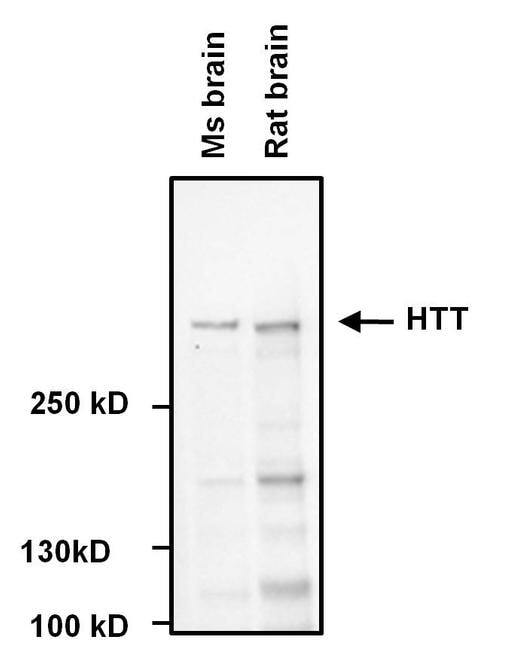
- MA3-040 has been used successfully in Western blotting to detect huntingtin in the brains lysates from mouse and rat.
- Reactivity
- Human, Mouse, Rat
- Host
- Mouse
- Isotype
- IgG
- Antibody clone number
- 1HU-4C8
- Vial size
- 50 µL
- Concentration
- Conc. Not Determined
- Storage
- -20° C, Avoid Freeze/Thaw Cycles
No comments: Submit comment
Supportive validation
- Submitted by
- Invitrogen Antibodies (provider)
- Main image

- Experimental details
- Western blot analysis of Huntingtin (HTT) was performed by loading the indicated whole cell lysates and 5 µL of PageRuler Plus Prestained Protein Ladder (Product # 26619) per well onto a Novex 4-20% Tris-Glycine polyacrylamide gel (Product # WT4202BOX ). Proteins were transferred to a nitrocellulose membrane using the G2 Blotter (Product # 62288), and blocked with Starting Block T20 (Product # 37543) for 1 hour at room temperature. HTT was detected at ~350 kD using a HTT monoclonal antibody (Product # MA3-040) at a dilution of 1:1000 in blocking buffer for 1 hour at room temperature on a rocking platform, followed by a Goat anti-Mouse IgG Secondary Antibody, HRP conjugate (Product # 31430) at a dilution of 1:20,000 for at least 30 minutes at room temperature. Chemiluminescent detection was performed using SuperSignal Pico substrate (Product # 34078) and the myECL Imager (Product # 62236).
- Submitted by
- Invitrogen Antibodies (provider)
- Main image
- Experimental details
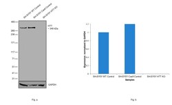
- Knockout of HTT was achieved by CRISPR-Cas9 genome editing using LentiArray™ Lentiviral sgRNA (Product # A32042, AssayID CRISPR1011411_LV) and LentiArray Cas9 Lentivirus (Product # A32064). Western blot analysis of HTT was performed by loading 20 µg of SH-SY5Y wild type (Lane 1), SH-SY5Y CAS9 (Lane 2), SH-SY5Y HTT KO (Lane 3) whole cell extracts. The blot was probed with Anti-Huntingtin Monoclonal Antibody (1HU-4C8)(Product # MA3-040) using 1:1000 dilution and Goat anti-Mouse IgG (H+L), Superclonal™ Recombinant Secondary Antibody, HRP (Product # A28177). Loss of signal upon CRISPR mediated knockout (KO) confirms that the antibody is specific to HTT.

- Submitted by
- Invitrogen Antibodies (provider)
- Main image
- Experimental details
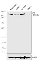
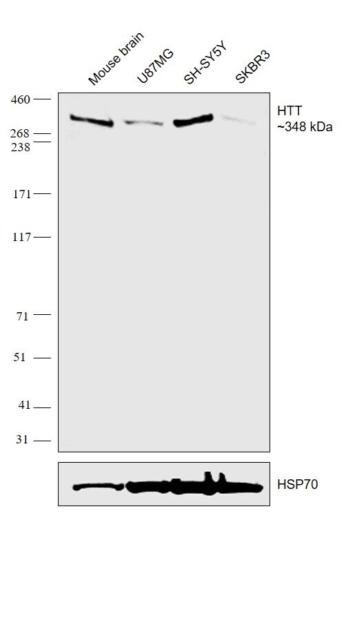
- Western blot was performed using Anti-Huntingtin Monoclonal Antibody (1HU-4C8)(Product # MA3-040) and a 348kDa band corresponding to Huntingtin was observed across the cell lines tested. Whole cell extracts (30 ug µg lysate) of Mouse Brain (Lane 1), U-87 MG (Lane 2), SH-SY5Y (Lane 3), SK-BR-3 (Lane 4) were electrophoresed using NuPAGE™ 3-8% Tris-Acetate Protein Gel (Product # EA0378BOX). Resolved proteins were then transferred onto a Nitrocellulose membrane (Product # LC2001) by iBlot® 2 Dry Blotting System (Product # IB21001). The blot was probed with the primary antibody (1:1000 dilution) and detected by chemiluminescence with Goat anti-Mouse IgG (H+L) Superclonal™ Recombinant Secondary Antibody, HRP (Product # A28177,1:4000 dilution) using the iBright FL 1000 (Product # A32752). Chemiluminescent detection was performed using Novex® ECL Chemiluminescent Substrate Reagent Kit (Product # WP20005).SK-BR-3 is expected to be a low HTT expressing model as reported.
Supportive validation
- Submitted by
- Invitrogen Antibodies (provider)
- Main image
- Experimental details
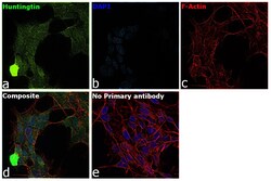
- Immunofluorescence analysis of Huntingtin was performed using 70% confluent log phase SH-SY5Y cells. The cells were fixed with 4% paraformaldehyde for 10 minutes, permeabilized with 0.1% Triton™ X-100 for 15 minutes, and blocked with 2% BSA for 45 minutes at room temperature. The cells were labeled with Huntingtin Monoclonal Antibody (1HU-4C8) (Product # MA3-040) at 1:200 dilution in 0.1% BSA, incubated at 4 degree celsius overnight and then labeled with Donkey anti-Mouse IgG (H+L) Highly Cross-Adsorbed Secondary Antibody, Alexa Fluor Plus 488 (Product # A32766), (1:2000 dilution), for 45 minutes at room temperature (Panel a: Green). Nuclei (Panel b:Blue) were stained with ProLong™ Diamond Antifade Mountant with DAPI (Product # P36962). F-actin (Panel c: Red) was stained with Rhodamine Phalloidin (Product # R415, 1:300 dilution). Panel d represents the merged image showing Cytoplasmic localization. Panel e represents control cells with no primary antibody to assess background. The images were captured at 60X with Oil immersion magnification.
