Explore
 Validate
 Learn
Learn Flow cytometry
Antibody data
- Antibody Data
- Antigen structure
- References [3]
- Comments [0]
- Validations
- Flow cytometry [2]
Submit
Validation data
Reference
Comment
Report error
- Product number
- FAB1418P - Provider product page

- Provider
- R&D Systems
- Product name
- Human Glut1 PE-conjugated Antibody
- Antibody type
- Monoclonal
- Description
- Protein A or G purified from hybridoma culture supernatant. Detects human Glut1. Stains human Glut1-transfected NS0 cells, but not NS0 control transfectants. Although Human Glut1 Antibody detects Glut1 on the surface of T cells (1, 2), it does not detect it on erythrocytes (5). The reason for this discrepancy is not understood, but may be related to conformational or post-translational modification differences.
- Reactivity
- Human
- Host
- Mouse
- Antigen sequence
AAA52571- Isotype
- IgG
- Antibody clone number
- 202915
- Vial size
- 100 Tests
- Storage
- Protect from light. Do not freeze. 12 months from date of receipt, 2 to 8 °C as supplied.
Submitted references IL-7 Restores T Lymphocyte Immunometabolic Failure in Septic Shock Patients through mTOR Activation.
Erosion of the chronic myeloid leukaemia stem cell pool by PPARγ agonists.
WASH knockout T cells demonstrate defective receptor trafficking, proliferation, and effector function.
Venet F, Demaret J, Blaise BJ, Rouget C, Girardot T, Idealisoa E, Rimmelé T, Mallet F, Lepape A, Textoris J, Monneret G
Journal of immunology (Baltimore, Md. : 1950) 2017 Sep 1;199(5):1606-1615
Journal of immunology (Baltimore, Md. : 1950) 2017 Sep 1;199(5):1606-1615
Erosion of the chronic myeloid leukaemia stem cell pool by PPARγ agonists.
Prost S, Relouzat F, Spentchian M, Ouzegdouh Y, Saliba J, Massonnet G, Beressi JP, Verhoeyen E, Raggueneau V, Maneglier B, Castaigne S, Chomienne C, Chrétien S, Rousselot P, Leboulch P
Nature 2015 Sep 17;525(7569):380-3
Nature 2015 Sep 17;525(7569):380-3
WASH knockout T cells demonstrate defective receptor trafficking, proliferation, and effector function.
Piotrowski JT, Gomez TS, Schoon RA, Mangalam AK, Billadeau DD
Molecular and cellular biology 2013 Mar;33(5):958-73
Molecular and cellular biology 2013 Mar;33(5):958-73
No comments: Submit comment
Supportive validation
- Submitted by
- R&D Systems (provider)
- Main image

- Experimental details
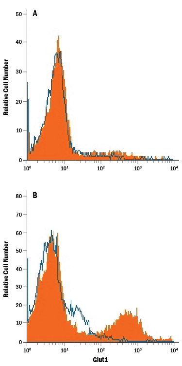
- Detection of Glut1 in Jurkat Human Cell Line by Flow Cytometry. Jurkat human acute T cell leukemia cell line either (A) untreated or (B) cultured in nutrient-depleted media was stained with Mouse Anti-Human Glut1 PE-conjugated Monoclonal Antibody (Catalog # FAB1418P, filled histogram) or isotype control antibody (Catalog # IC0041P, open histogram). View our protocol for Staining Membrane-associated Proteins.
- Submitted by
- R&D Systems (provider)
- Main image
- Experimental details
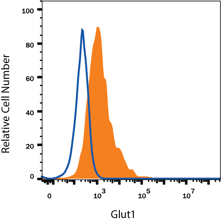
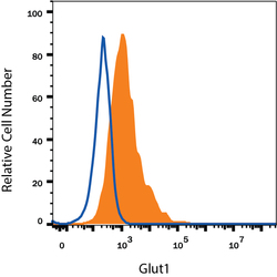
- Detection of Glut1 in HepG2 Human Cell Line by Flow Cytometry. HepG2 human hepatocellular carcinoma cell line was stained with Mouse Anti-Human Glut1 PE-conjugated Monoclonal Antibody (Catalog # FAB1418P, filled histogram) or isotype control antibody (Catalog # IC0041P, open histogram). View our protocol for Staining Membrane-associated Proteins.