Explore
 Validate
 Learn
LearnMA5-16167
antibody from Invitrogen Antibodies
Targeting: TP73
P73
 Western blot Immunocytochemistry
 Immunoprecipitation Immunohistochemistry Flow cytometry Chromatin Immunoprecipitation
Antibody data
- Antibody Data
- Antigen structure
- References [0]
- Comments [0]
- Validations
- Western blot [4]
- Immunocytochemistry [1]
- Immunohistochemistry [1]
- Flow cytometry [2]
Submit
Validation data
Reference
Comment
Report error
- Product number
- MA5-16167 - Provider product page

- Provider
- Invitrogen Antibodies
- Product name
- p73 Monoclonal Antibody (5B429)
- Antibody type
- Monoclonal
- Antigen
- Other
- Reactivity
- Human, Mouse, Rat
- Host
- Mouse
- Isotype
- IgG
- Antibody clone number
- 5B429
- Vial size
- 100 µg
- Concentration
- 1.0 mg/mL
- Storage
- Store at 4°C short term. For long term storage, store at -20°C, avoiding freeze/thaw cycles.
No comments: Submit comment
Supportive validation
- Submitted by
- Invitrogen Antibodies (provider)
- Main image

- Experimental details
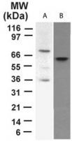
- Western blot analysisof p73 in A) transfectedcell lysate and B) HeLa cell lysate using a p73 monoclonal antibody (Product # MA5-16167) at 2 µg/mL.
- Submitted by
- Invitrogen Antibodies (provider)
- Main image
- Experimental details
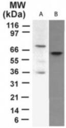
- Western blot analysis of p73 in A) transfected cell lysate and B) HeLa cell lysate. Samples were incubated in p73 monoclonal antibody (Product # MA5-16167) using a dilution of 2 µg/mL.

- Submitted by
- Invitrogen Antibodies (provider)
- Main image

- Experimental details
- Western blot analysis of p73 in 1.0 mg/mL HeLa lysate. Samples were incubated in p73 monoclonal (Product # MA5-16167). This experiment was performed under reducing conditions using the 12-230 kDa separation system. * Non-specific interaction with the 230 kDa Simple Western standard may be seen with this antibody.

- Submitted by
- Invitrogen Antibodies (provider)
- Main image

- Experimental details
- Western blot analysis was performed on modified whole cell extracts (1% SDS) (30 µg lysate) of HeLa (Lane 1), SK-Ov-3 (Lane 2), SK-BR-3 (Lane 3), MCF-7 (Lane 4) and MDA-MB-231 (Lane 5). The blot was probed with p73 Monoclonal Antibody (5B429) (Product # MA5-16167, 1µg/ml) and detected by chemiluminescence using Goat anti-Mouse IgG (H+L) Superclonal™ Secondary Antibody, HRP conjugate (Product # A28177, 0.25µg/ml, 1:4000 dilution). A 40kDa band corresponding to P73 was observed across the cell lines tested.

Supportive validation
- Submitted by
- Invitrogen Antibodies (provider)
- Main image

- Experimental details
- Immunocytochemistry analysis of p73 in immersion fixed Hela human cell line using. Samples were incubated in p73 monoclonal antibody (Product # MA5-16167) using a dilution of 3 µg/mL for 3 hours at room temperature followed by NorthernLights™ 557-conjugated Anti-Mouse IgG Secondary Antibody (red) and counterstained with DAPI (blue). Staining was observed in the cytoplasm and in the nuclei.

Supportive validation
- Submitted by
- Invitrogen Antibodies (provider)
- Main image

- Experimental details
- Immunohistochemical analysis of p73 in formalin-fixed paraffin-embedded human breast cancer tissue. Samples were incubated in p73 monoclonal antibody (Product # MA5-16167) using a dilution of 5 µg/mL. Staining of tissues is enhanced by boiling tissue sections in 10 mM sodium citrate buffer, pH 6.0 for 10-20 min followed by cooling at RT for 20 min.

Supportive validation
- Submitted by
- Invitrogen Antibodies (provider)
- Main image
- Experimental details
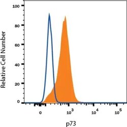
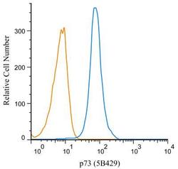
- Flow cytometry of p73 in Human HeLa Cell Line. Samples were incubated in p73 monoclonal antibody (Product # MA5-16167) or Mouse IgG1 isotype control followed by APC-conjugated Anti-Mouse IgG Secondary Antibody. To facilitate intracellular staining, cells were fixed with Flow Cytometry Fixation Buffer and permeabilized with Flow Cytometry Permeabilization/Wash Buffer.

- Submitted by
- Invitrogen Antibodies (provider)
- Main image
- Experimental details
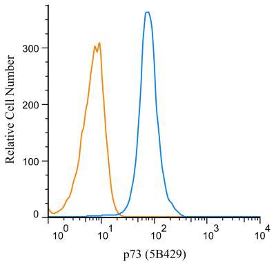
- Flow cytometry of p73 in HeLa cells. Samples were incubated in p73 monoclonal antibody (Product # MA5-16167) using a dilution of 1 µg/mL for 30 minutes followed by mouse F(ab)2 IgG (H+L) APC-conjugated secondary antibody. Antibody (blue) and a matched isotype control (orange). Cells were fixed with 4% PFA and then permeablized with 0.1% saponin.