 Explore
 Validate
 Learn
Learn Western blot
 Immunoprecipitation
Antibody data
- Antibody Data
- Antigen structure
- References [0]
- Comments [0]
- Validations
- Western blot [4]
- Immunohistochemistry [2]
Submit
Validation data
Reference
Comment
Report error
- Product number
- MA5-16168 - Provider product page

- Provider
- Invitrogen Antibodies
- Product name
- p73 Monoclonal Antibody (5B1288)
- Antibody type
- Monoclonal
- Antigen
- Other
- Description
- Suggested positive control: antigen standard for TP73 (transient overexpression lysate), p73 transfected cells..
- Reactivity
- Human, Mouse
- Host
- Mouse
- Isotype
- IgG
- Antibody clone number
- 5B1288
- Vial size
- 100 µg
- Concentration
- 1 mg/mL
- Storage
- -20° C, Avoid Freeze/Thaw Cycles
No comments: Submit comment
Supportive validation
- Submitted by
- Invitrogen Antibodies (provider)
- Main image

- Experimental details
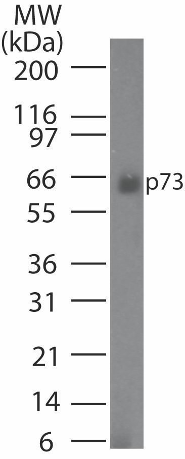
- Western blot analysis of p73 in HeLa cell lysate using a p73 (Alpha, Beta, Gamma, Delta Isoforms) monoclonal antibody (Product # MA5-16168) at1 µg/mL.
- Submitted by
- Invitrogen Antibodies (provider)
- Main image

- Experimental details
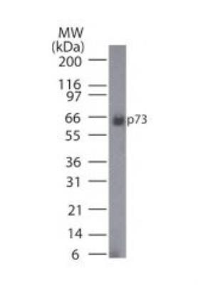
- Western blot analysis of p73 in HeLa cell lysate. Samples were incubated in p73 monoclonal antibody (Product # MA5-16168) using a dilution of 1 µg/mL.
- Submitted by
- Invitrogen Antibodies (provider)
- Main image
- Experimental details
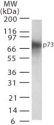
- Western blot analysis of p73 in transfected cell lysate. Sample was incubated in p73 monoclonal antibody (Product # MA5-16168).
- Submitted by
- Invitrogen Antibodies (provider)
- Main image
- Experimental details
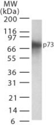
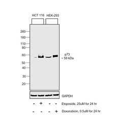
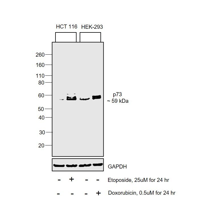
- Western blot was performed using Anti-p73 Pan Monoclonal Antibody (5B1288) (Product # MA5-16168) and a 59 kDa band corresponding to one of the isoform of Tumor protein p73 was observed in HCT 116 upon treatment with Etoposide. The expression of protein was also increased in HEK-293 upon Doxorubicin treatment. Nuclear enriched extracts (30 µg lysate) of HCT 116 (Lane 1) and HCT 116 treated with Etoposide (25 µM for 24hr) (Lane 2), HEK-293 (Lane 3) and HEK-293 treated with Doxorubicin (0.5 µM for 24hr) (Lane 4) were electrophoresed using NuPAGE™ 4-12% Bis-Tris Protein Gel (Product # NP0322BOX). Resolved proteins were then transferred onto a Nitrocellulose membrane (Product # IB23001) by iBlot® 2 Dry Blotting System (Product # IB21001). The blot was probed with the primary antibody (1 µg/mL) and detected by chemiluminescence with Goat anti-Mouse IgG (H+L) Superclonal™ Recombinant Secondary Antibody, HRP (Product # A28177, 1:4000 dilution) using the iBright FL 1000 (Product # A32752). Chemiluminescent detection was performed using SuperSignal™ West Dura Extended Duration Substrate (Product # 34076).
Supportive validation
- Submitted by
- Invitrogen Antibodies (provider)
- Main image

- Experimental details
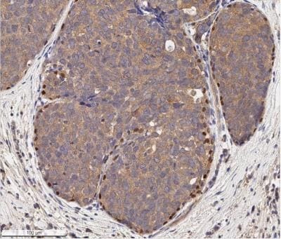
- Immunohistochemical analysis of p73 in Human breast cancer section. Samples were incubated in p73 monoclonal antibody (Product # MA5-16168) using a dilution of 10 µg/mL. Bond Rx autostainer (Leica Biosystems). The assay involved 20 minutes of heat induced antigen retrieval (HIER) with 10 mM sodium citrate buffer (pH 6.0) and endogenous peroxidase quenching using peroxide block. The sections were incubated with primary antibody for 30 minutes. Bond Polymer Refine Detection (Leica Biosystems) and DAB were used for signal detection which followed counterstaining with hematoxylin. Whole slide scanning and capturing of representative images (20X) were performed using Aperio AT2 (Leica Biosystems). The cancer cells showed cytoplasmic immunoreactivity for p73 and the signal was very weak in the core and stroma of tumors. Peripheral cells of tumor areas, apparently the myoepithelial cells, showed a strong nuclear staining of p73.
- Submitted by
- Invitrogen Antibodies (provider)
- Main image
- Experimental details
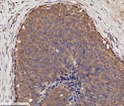
- Immunohistochemical analysis of p73 in Human breast cancer section. Samples were incubated in p73 monoclonal antibody (Product # MA5-16168) using a dilution of 10 µg/mL. Bond Rx autostainer (Leica Biosystems). The assay involved 20 minutes of heat induced antigen retrieval (HIER) with 10 mM sodium citrate buffer (pH 6.0) and endogenous peroxidase quenching using peroxide block. The sections were incubated with primary antibody for 30 minutes. Bond Polymer Refine Detection (Leica Biosystems) and DAB were used for signal detection which followed counterstaining with hematoxylin. Whole slide scanning and capturing of representative images (20X) were performed using Aperio AT2 (Leica Biosystems). The cancer cells showed cytoplasmic immunoreactivity for p73 and the signal was very weak in the core and stroma of tumors. Peripheral cells of tumor areas, apparently the myoepithelial cells, showed a strong nuclear staining of p73.
