Explore
 Validate
 Learn
Learn Western blot
Antibody data
- Antibody Data
- Antigen structure
- References [1]
- Comments [0]
- Validations
- Western blot [3]
- Immunohistochemistry [1]
Submit
Validation data
Reference
Comment
Report error
- Product number
- 700685 - Provider product page

- Provider
- Invitrogen Antibodies
- Product name
- NMDAR1 Recombinant Rabbit Monoclonal Antibody (1H13L3)
- Antibody type
- Monoclonal
- Antigen
- Recombinant full-length protein
- Description
- This antibody is predicted to react with mouse and rat based on sequence homology.
- Antibody clone number
- 1H13L3
- Concentration
- 0.5 mg/mL
Submitted references Stereotactic injection of cerebrospinal fluid from anti-NMDA receptor encephalitis into rat dentate gyrus impairs NMDA receptor function.
Würdemann T, Kersten M, Tokay T, Guli X, Kober M, Rohde M, Porath K, Sellmann T, Bien CG, Köhling R, Kirschstein T
Brain research 2016 Feb 15;1633:10-18
Brain research 2016 Feb 15;1633:10-18
No comments: Submit comment
Supportive validation
- Submitted by
- Invitrogen Antibodies (provider)
- Main image
- Experimental details
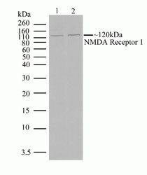
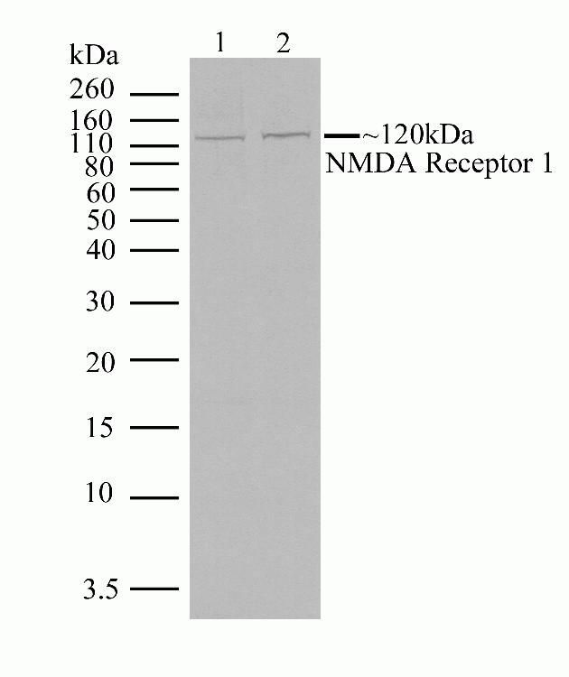
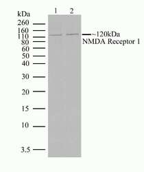
- Western blot analysis of NMDA Receptor 1 in rat brain lysate (30 µg/lane) (lane 1) and mouse brain lysate (30 µg/lane) (lane 2) using a NMDA Receptor 1 recombinant rabbit monoclonal antibody (Product # 700685) at a dilution of 2.5 µg/mL. NBT/BCIP was used as the substrate (Product # WB7105).
- Submitted by
- Invitrogen Antibodies (provider)
- Main image

- Experimental details
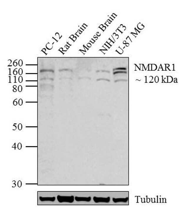
- Western blot analysis of NMDAR1 was performed by loading 20 µg of PC-12 (lane1), Rat Brain (lane2), Mouse Brain (lane3), NIH/3T3 (lane4) and U-87 MG (lane5) lysates using Novex®NuPAGE®4-12% Bis-Tris gel (Product # NP0321BOX), XCell SureLock Electrophoresis System (Product # EI0002), Novex® Sharp Pre-Stained Protein Standard (Product # LC5800). Proteins were transferred to a PVDF membrane and blocked with 5% skim milk for 1 hour at room temperature. NMDAR1 was detected at ~120 kDa using NMDAR1 Recombinant Rabbit Monoclonal Antibody (Product # 700685) at 1-2 µg/mL in 2.5% skim milk at 4°C overnight on a rocking platform. Goat anti-Rabbit IgG - HRP Secondary Antibody (Product # G-21234) at 1:5000 dilution was used and chemiluminescent detection was performed using Pierce™ ECL Western blotting Substrate (Product # 32106).
- Submitted by
- Invitrogen Antibodies (provider)
- Main image
- Experimental details
- Western blot analysis of NMDA Receptor 1 in rat brain lysate (30 µg/lane) (lane 1) and mouse brain lysate (30 µg/lane) (lane 2) using a NMDA Receptor 1 recombinant rabbit monoclonal antibody (Product # 700685) at a dilution of 2.5 µg/mL. NBT/BCIP was used as the substrate (Product # WB7105).

Supportive validation
- Submitted by
- Invitrogen Antibodies (provider)
- Main image
- Experimental details
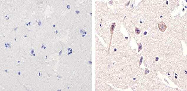
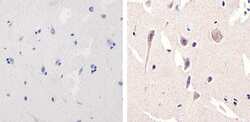
- Immunohistochemistry analysis of NMDAR1 showing staining in the cytoplasm of paraffin-embedded human brain tissue (right) compared to a negative control without primary antibody (left). To expose target proteins, antigen retrieval was performed using 10mM sodium citrate (pH 6.0), microwaved for 8-15 min. Following antigen retrieval, tissues were blocked in 3% H2O2-methanol for 15 min at room temperature, washed with ddH2O and PBS, and then probed with a NMDAR1 monoclonal antibody (Product # 700685) diluted in 3% BSA-PBS at a dilution of 1:20 overnight at 4°C in a humidified chamber. Tissues were washed extensively in PBST and detection was performed using an HRP-conjugated secondary antibody followed by colorimetric detection using a DAB kit. Tissues were counterstained with hematoxylin and dehydrated with ethanol and xylene to prep for mounting.