Explore
 Validate
 Learn
Learn Western blot
Antibody data
- Antibody Data
- Antigen structure
- References [0]
- Comments [0]
- Validations
- Western blot [2]
- Immunohistochemistry [1]
Submit
Validation data
Reference
Comment
Report error
- Product number
- 710020 - Provider product page

- Provider
- Invitrogen Antibodies
- Product name
- NMDAR1 Recombinant Polyclonal Antibody (1HCLC)
- Antibody type
- Polyclonal
- Antigen
- Recombinant full-length protein
- Description
- This antibody is predicted to react with mouse based on sequence homology.
- Antibody clone number
- 1HCLC
- Concentration
- 0.5 mg/mL
No comments: Submit comment
Supportive validation
- Submitted by
- Invitrogen Antibodies (provider)
- Main image
- Experimental details
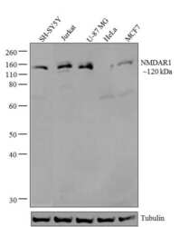
- Western blot analysis of NMDAR1 was performed by loading 20 µg of SH-SY5Y (lane1), Jurkat (lane2), U-87 MG (lane3), HeLa (lane4) and MCF7 (lane5) cell lysates using Novex®NuPAGE®4-12 % Bis-Tris gel (Product # NP0321BOX), XCell SureLock Electrophoresis System (Product # EI0002), Novex® Sharp Pre-Stained Protein Standard (Product # LC5800). Proteins were transferred to a PVDF membrane and blocked with 5 % skim milk for 1 hour at room temperature. NMDAR1 was detected at ~120 kDa using NMDAR1 Recombinant Rabbit Polyclonal Antibody (Product # 710020) at 1-3 µg/mL in 2.5 % skim milk at 4°C overnight on a rocking platform. Goat anti-Rabbit IgG-HRP Secondary Antibody (Product # G-21234) at 1:5000 dilution was used and chemiluminescent detection was performed using Pierce™ ECL Western blotting Substrate (Product # 32106).

- Submitted by
- Invitrogen Antibodies (provider)
- Main image

- Experimental details
- Western blot analysis of NMDA Receptor 1 in rat brain lysate (30 µg/lane) (lane 1) and mouse brain lysate (30 µg/lane) (lane 2) using a NMDA Receptor 1 Recombinant Rabbit Polyclonal Antibody (Product # 710020) at a dilution of 5 µg/mL. NBT/BCIP was used as the substrate (Product # WB7105).

Supportive validation
- Submitted by
- Invitrogen Antibodies (provider)
- Main image

- Experimental details
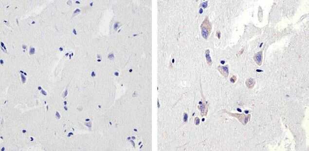
- Immunohistochemistry analysis of NMDAR1 showing staining in the cytoplasm of paraffin-embedded human brain tissue (right) compared to a negative control without primary antibody (left). To expose target proteins, antigen retrieval was performed using 10mM sodium citrate (pH 6.0), microwaved for 8-15 min. Following antigen retrieval, tissues were blocked in 3% H2O2-methanol for 15 min at room temperature, washed with ddH2O and PBS, and then probed with a NMDAR1 Recombinant Rabbit Polyclonal Antibody (Product # 710020) diluted in 3% BSA-PBS at a dilution of 1:20 overnight at 4°C in a humidified chamber. Tissues were washed extensively in PBST and detection was performed using an HRP-conjugated secondary antibody followed by colorimetric detection using a DAB kit. Tissues were counterstained with hematoxylin and dehydrated with ethanol and xylene to prep for mounting.