 Explore
 Validate
 Learn
Learn Immunocytochemistry
Antibody data
- Antibody Data
- Antigen structure
- References [1]
- Comments [0]
- Validations
- Immunocytochemistry [2]
- Other assay [1]
Submit
Validation data
Reference
Comment
Report error
- Product number
- MA1-014-D550 - Provider product page

- Provider
- Invitrogen Antibodies
- Product name
- SOX2 Monoclonal Antibody (20G5), DyLight™ 550
- Antibody type
- Monoclonal
- Antigen
- Recombinant full-length protein
- Description
- MA1-014-D550 has been successfully used in ICC/IF applications with human and mouse samples.
- Reactivity
- Human, Mouse
- Host
- Mouse
- Conjugate
- Yellow dye
- Isotype
- IgG
- Antibody clone number
- 20G5
- Vial size
- 100 µL
- Concentration
- 1 mg/mL
- Storage
- 4° C, do not freeze
Submitted references Recessive Inheritance of Congenital Hydrocephalus With Other Structural Brain Abnormalities Caused by Compound Heterozygous Mutations in ATP1A3.
Allocco AA, Jin SC, Duy PQ, Furey CG, Zeng X, Dong W, Nelson-Williams C, Karimy JK, DeSpenza T, Hao LT, Reeves B, Haider S, Gunel M, Lifton RP, Kahle KT
Frontiers in cellular neuroscience 2019;13:425
Frontiers in cellular neuroscience 2019;13:425
No comments: Submit comment
Supportive validation
- Submitted by
- Invitrogen Antibodies (provider)
- Main image
- Experimental details
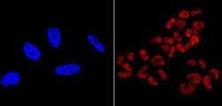
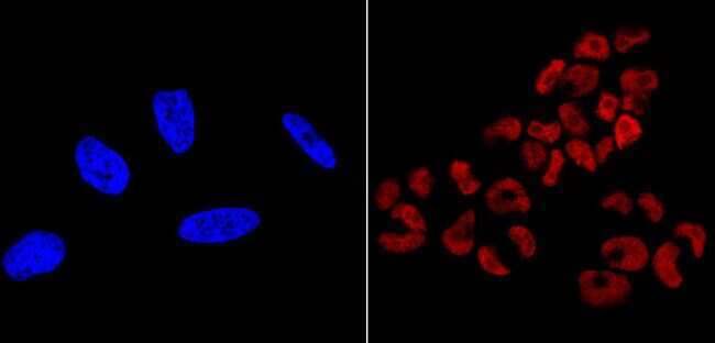
- Immunofluorescent analysis of SOX2 (red) showing nuclear staining of NCCIT cells (right panel) compared to negative HeLa cell control (left panel). The cells were fixed with formalin for 15 minutes, permeabilized with 0.1% Triton X-100 in TBS, washed, and then blocked with 3% BSA-PBS for 30 minutes at room temperature. Cells were probed with a DyLight 550-conjugated SOX2 monoclonal antibody (Product # MA1-014-D550) in 3% BSA-PBS at a dilution of 1:50 and incubated for 1 hour at 37C in the dark. Nuclei (left panel, blue) were stained with DAPI. Images were taken at 60X magnification.
- Submitted by
- Invitrogen Antibodies (provider)
- Main image
- Experimental details
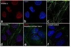
- Immunofluorescence analysis of SOX2 was performed using 70% confluent log phase NTERA-2 cells. The cells were fixed with 4% paraformaldehyde for 10 minutes, permeabilized with 0.1% Triton™ X-100 for 15 minutes, and blocked with 1% BSA for 1 hour at room temperature. The cells were labeled with SOX2 Mouse Monoclonal Antibody (Product # MA1-014-D550) at 5 µg/mL in 0.1% BSA, incubated at 4 degree celsius overnight (Panel a: red). Nuclei (Panel b: blue) were stained with ProLong™ Diamond Antifade Mountant with DAPI (Product # P36962). F-actin (Panel c: green) was stained with Alexa Fluor™ 488 Phalloidin (Product # A12379, 1:300). Panel d represents the merged image showing nuclear localization. Panel e shows SOX2 negative cell line HeLa with no signal. Panel f represents control cells with Isotype control to assess background. The images were captured at 60X magnification.

Supportive validation
- Submitted by
- Invitrogen Antibodies (provider)
- Main image
- Experimental details
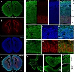
- FIGURE 2 Expression of Atp1a3 in the embryonic mouse brain at E15.5. Fluorescent images taken at low magnification showing expression of (A) Atp1a3, (B) Sox2, (C) NeuN, and (D) all channels merged in the embryonic mouse brain. Dotted boxes in panel A identify the cortex and the choroid plexus. High magnification cortical expression of (E) Atp1a3, (F) Sox2, (G) NeuN, and (H) all channels merged. MZ - marginal zone, CP - cortical plate, IZ - intermediate zone, SVZ - subventricular zone, VZ - ventricular zone. Dotted boxes in panel (E) shows the cortical plate and ventricular zone. (I-K) High magnification expression of Atp1a3 in NeuN + cells in the CP. (L-N) Expression of Atp1a3 in Sox2 + cells in the ventricular zone. (O,P) High-magnification expression of Atp1a3 in the choroid plexus. (Q) Brain sections incubated with Atp1a3 primary antibody alone or (R) Atp1a3 primary antibody blocked with immunizing peptide.
