Explore
 Validate
 Learn
Learn Immunocytochemistry
 Immunoprecipitation
Antibody data
- Antibody Data
- Antigen structure
- References [4]
- Comments [0]
- Validations
- Immunocytochemistry [1]
- Flow cytometry [3]
Submit
Validation data
Reference
Comment
Report error
- Product number
- MA1-10135 - Provider product page

- Provider
- Invitrogen Antibodies
- Product name
- CD20 Monoclonal Antibody (2H7)
- Antibody type
- Monoclonal
- Antigen
- Other
- Reactivity
- Human
- Host
- Mouse
- Isotype
- IgG
- Antibody clone number
- 2H7
- Vial size
- 100 µg
- Concentration
- 1.0 mg/mL
- Storage
- 4° C, do not freeze
Submitted references Longitudinal analyses reveal distinct immune response landscapes in lung and intestinal tissues from SARS-CoV-2-infected rhesus macaques.
Mucosal Inducible NO Synthase-Producing IgA+ Plasma Cells in Helicobacter pylori-Infected Patients.
Effects of B Cell Depletion on Early Mycobacterium tuberculosis Infection in Cynomolgus Macaques.
Evidence for innate immune system activation in HIV type 1-infected elite controllers.
Zheng H, Chen Y, Li J, Li H, Zhao X, Li J, Yang F, Li Y, Liu C, Qin L, Zuo Y, Zhang Q, He Z, Shi H, Li Q, Liu L
Cell reports 2022 May 24;39(8):110864
Cell reports 2022 May 24;39(8):110864
Mucosal Inducible NO Synthase-Producing IgA+ Plasma Cells in Helicobacter pylori-Infected Patients.
Neumann L, Mueller M, Moos V, Heller F, Meyer TF, Loddenkemper C, Bojarski C, Fehlings M, Doerner T, Allers K, Aebischer T, Ignatius R, Schneider T
Journal of immunology (Baltimore, Md. : 1950) 2016 Sep 1;197(5):1801-8
Journal of immunology (Baltimore, Md. : 1950) 2016 Sep 1;197(5):1801-8
Effects of B Cell Depletion on Early Mycobacterium tuberculosis Infection in Cynomolgus Macaques.
Phuah J, Wong EA, Gideon HP, Maiello P, Coleman MT, Hendricks MR, Ruden R, Cirrincione LR, Chan J, Lin PL, Flynn JL
Infection and immunity 2016 May;84(5):1301-1311
Infection and immunity 2016 May;84(5):1301-1311
Evidence for innate immune system activation in HIV type 1-infected elite controllers.
Krishnan S, Wilson EM, Sheikh V, Rupert A, Mendoza D, Yang J, Lempicki R, Migueles SA, Sereti I
The Journal of infectious diseases 2014 Mar;209(6):931-9
The Journal of infectious diseases 2014 Mar;209(6):931-9
No comments: Submit comment
Supportive validation
- Submitted by
- Invitrogen Antibodies (provider)
- Main image
- Experimental details
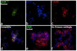
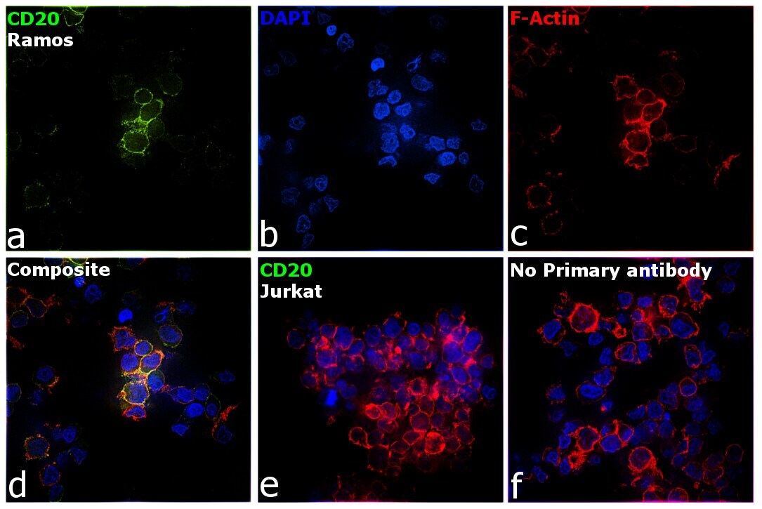
- Immunofluorescence analysis of CD20 was performed using 70% confluent log phase Ramos cells. The cells were fixed with 4% paraformaldehyde for 5 minutes, permeabilized with 0.1% Triton™ X-100 for 10 minutes, and blocked with 2% BSA for 45 minutes at room temperature. The cells were labeled with CD20 Monoclonal Antibody (2H7) (Product # MA1-10135) at 5 µg/mL concentration in 0.1% BSA, incubated at 4 degree celsius overnight and then labeled with Donkey anti-Mouse IgG (H+L) Highly Cross-Adsorbed Secondary Antibody, Alexa Fluor Plus 488 (Product # A32766), (1:2000 dilution), for 45 minutes at room temperature (Panel a: Green). Nuclei (Panel b: Blue) were stained with ProLong™ Diamond Antifade Mountant with DAPI (Product # P36962). F-actin (Panel c: Red) was stained with Rhodamine Phalloidin (Product # R415, 1:300). Panel d represents the merged image showing plasma membrane and cytoplasm localization. Panel e represents negative cell line Jurkat. Panel f represents control cells with no primary antibody to assess background. The images were captured at 60X magnification.
Supportive validation
- Submitted by
- Invitrogen Antibodies (provider)
- Main image
- Experimental details
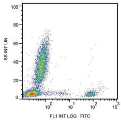
- Flow cytometry surface staining of human peripheral blood with anti-CD20 monoclonal antibody (Product # MA1-10135).
- Submitted by
- Invitrogen Antibodies (provider)
- Main image
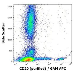
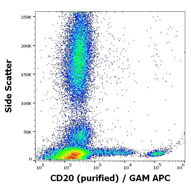
- Experimental details
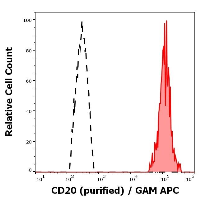
- Flow cytometry surface staining pattern of human peripheral whole blood stained using anti-human CD20 (2H7) purified Monoclonal antibody (Product # MA1-10135); concentration in sample 0.6 µg/mL, GAM APC.
- Submitted by
- Invitrogen Antibodies (provider)
- Main image

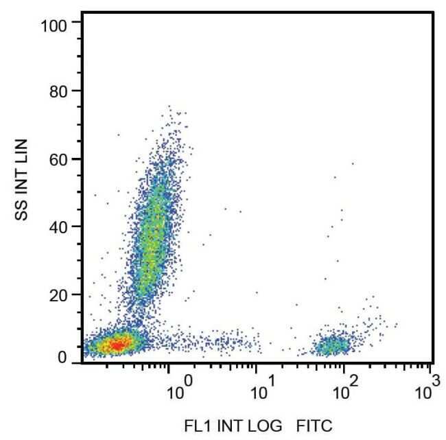
- Experimental details
- Separation of human CD20 positive lymphocytes (red-filled) from neutrofil granulocytes (black-dashed) in flow cytometry analysis (surface staining) of peripheral whole blood stained using anti-human CD20 (2H7) purified Monoclonal antibody (Product # MA1-10135); concentration in sample 0.6 µg/mL, GAM APC.