Explore
 Validate
 Learn
Learn Western blot
Antibody data
- Antibody Data
- Antigen structure
- References [4]
- Comments [0]
- Validations
- Western blot [3]
- Immunohistochemistry [1]
- Flow cytometry [1]
- Other assay [1]
Submit
Validation data
Reference
Comment
Report error
- Product number
- PA1-9024 - Provider product page

- Provider
- Invitrogen Antibodies
- Product name
- CD20 Polyclonal Antibody
- Antibody type
- Polyclonal
- Antigen
- Synthetic peptide
- Reactivity
- Human
- Host
- Goat
- Isotype
- IgG
- Vial size
- 100 µg
- Concentration
- 0.5 mg/mL
- Storage
- -20° C, Avoid Freeze/Thaw Cycles
Submitted references RELT stains prominently in B-cell lymphomas and binds the hematopoietic transcription factor MDFIC.
Memory B-cell aggregates in skin biopsy are diagnostic for primary Sjögren's syndrome.
BAFF-modulated repopulation of B lymphocytes in the blood and salivary glands of rituximab-treated patients with Sjögren's syndrome.
Identification of transitional type II B cells in the salivary glands of patients with Sjögren's syndrome.
Cusick JK, Alhomsy Y, Wong S, Talbott G, Uversky VN, Hart C, Hejazi N, Jacobs AT, Shi Y
Biochemistry and biophysics reports 2020 Dec;24:100868
Biochemistry and biophysics reports 2020 Dec;24:100868
Memory B-cell aggregates in skin biopsy are diagnostic for primary Sjögren's syndrome.
Roguedas AM, Pers JO, Lemasson G, Devauchelle V, Tobón GJ, Saraux A, Misery L, Youinou P
Journal of autoimmunity 2010 Nov;35(3):241-7
Journal of autoimmunity 2010 Nov;35(3):241-7
BAFF-modulated repopulation of B lymphocytes in the blood and salivary glands of rituximab-treated patients with Sjögren's syndrome.
Pers JO, Devauchelle V, Daridon C, Bendaoud B, Le Berre R, Bordron A, Hutin P, Renaudineau Y, Dueymes M, Loisel S, Berthou C, Saraux A, Youinou P
Arthritis and rheumatism 2007 May;56(5):1464-77
Arthritis and rheumatism 2007 May;56(5):1464-77
Identification of transitional type II B cells in the salivary glands of patients with Sjögren's syndrome.
Daridon C, Pers JO, Devauchelle V, Martins-Carvalho C, Hutin P, Pennec YL, Saraux A, Youinou P
Arthritis and rheumatism 2006 Jul;54(7):2280-8
Arthritis and rheumatism 2006 Jul;54(7):2280-8
No comments: Submit comment
Supportive validation
- Submitted by
- Invitrogen Antibodies (provider)
- Main image
- Experimental details
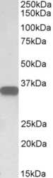
- Western blot analysis of CD20 by a CD20 monoclonal antibody (Product # PA1-9024) at a concentration of 0.05 µg/mL. Human Lymph Node lysate (35µg protein in RIPA buffer). Detected by chemiluminescence.

- Submitted by
- Invitrogen Antibodies (provider)
- Main image

- Experimental details
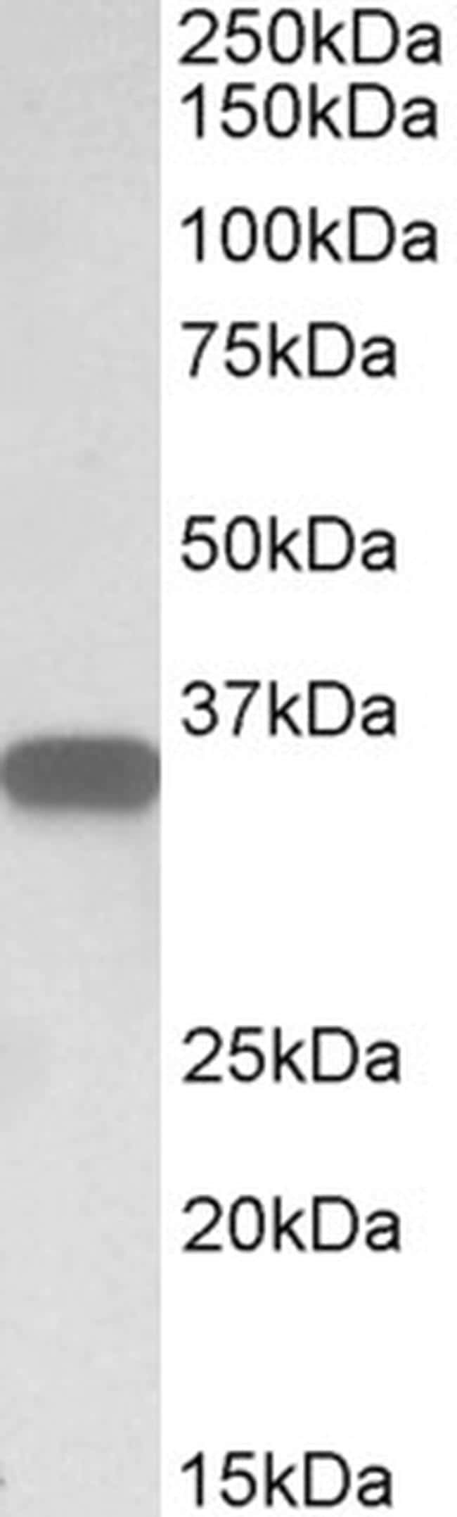
- Western blot detection of MS4A1 in Daudi lysate using Product # PA1-9024.
- Submitted by
- Invitrogen Antibodies (provider)
- Main image

- Experimental details
- Western blot analysis of CD20 by a CD20 monoclonal antibody (Product # PA1-9024) at a concentration of 0.05 µg/mL. Human Lymph Node lysate (35µg protein in RIPA buffer). Detected by chemiluminescence.
Supportive validation
- Submitted by
- Invitrogen Antibodies (provider)
- Main image
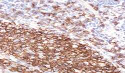
- Experimental details
- Immunohistochemical analysis of CD20 in Human Tonsil using a CD20 monoclonal antibody (Product #PA1-9024) at 2 µg/mL. The Human Tonsil tissue section was paraffin embeded and detected using steamed antigen retrieval with citrate buffer pH 6, HRP-staining.

Supportive validation
- Submitted by
- Invitrogen Antibodies (provider)
- Main image
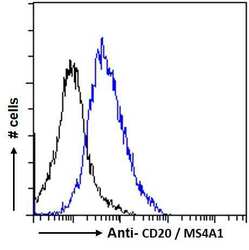
- Experimental details
- Flow cytometric analysis of CD20 in Daudi cells using a polyclonal antibody (Product #PA1-9024). Daudi cells (blue line) were paraformaldehyde fixed and permeabilized with 0.5% Triton. The primary antibody was incubated for one hour (10 µg/mL) followed by an Alexa Fluor 488 secondary antibody (1 µg/mL). IgG control: Unimmunized goat IgG (black line) followed by an Alexa Fluor 488 secondary antibody.

Supportive validation
- Submitted by
- Invitrogen Antibodies (provider)
- Main image
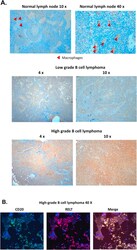
- Experimental details
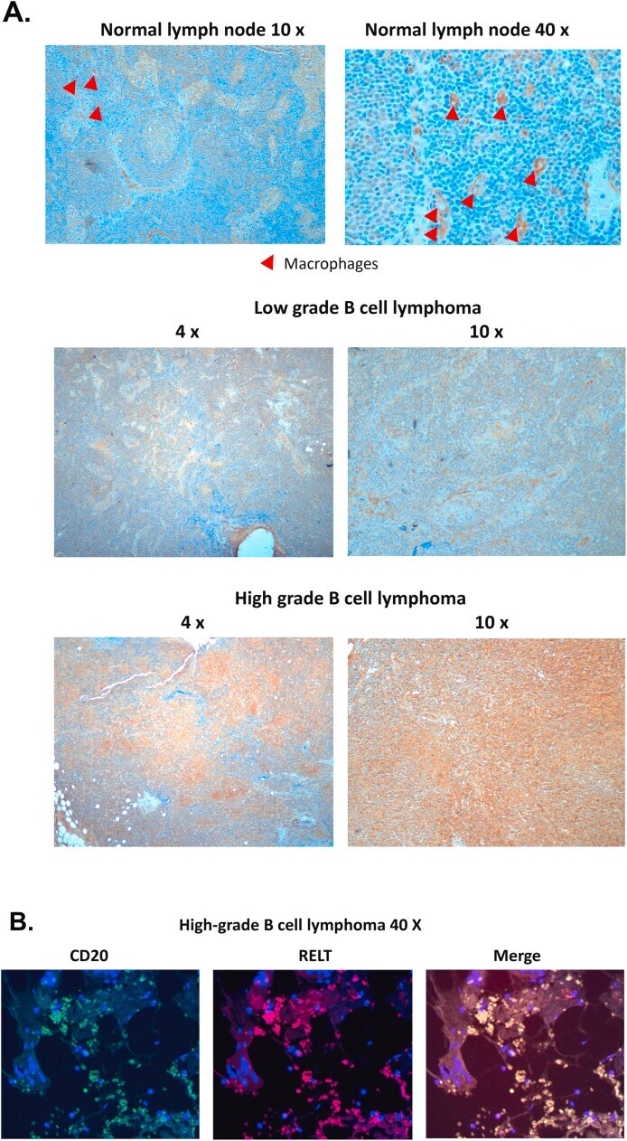
- Fig. 5 RELT is expressed in human lymph nodes and B-cell lymphomas. Immunohistochemistry was performed on both normal lymph nodes and resected B-cell lymphomas using an antibody directed against RELT as described in Materials and Methods. (A) Comparison of RELT staining in normal lymph nodes, low-grade, and high-grade B-cell lymphomas. RELT staining at the indicated magnification with the presence of macrophages in normal lymph nodes indicated by red arrowheads. (B) RELT co-localizes with CD20 in B-cell lymphomas. Immunofluorescence was conducted with RELT and CD20 antibodies as described in Materials and Methods resulting in RELT fluorescing red, CD20 fluorescing green, and DAPI staining nuclei blue. Photographs were taken at 40x magnification. (For interpretation of the references to color in this figure legend, the reader is referred to the Web version of this article.) Fig. 5