 Explore
 Validate
 Learn
Learn Western blot
 Immunohistochemistry
Antibody data
- Antibody Data
- Antigen structure
- References [2]
- Comments [0]
- Validations
- Western blot [1]
- Immunocytochemistry [4]
Submit
Validation data
Reference
Comment
Report error
- Product number
- GTX27260 - Provider product page

- Provider
- GeneTex
- Proper citation
- GeneTex Cat#GTX27260, RRID:AB_370784
- Product name
- GFAP antibody
- Antibody type
- Polyclonal
- Reactivity
- Human, Mouse, Rat, Bovine, Canine, Chicken/Avian, Feline, Guinea Pig, Porcine, Rabbit, Zebrafish
- Host
- Rabbit
Submitted references Peripheral nerve sheath tumor invading the nasal cavities of a 6-year-old female Pointer dog.
Spontaneous schwannoma in zebrafish, Danio rerio (Hamilton).
Sfacteria A, Perillo L, Macrì F, Lanteri G, Rifici C, Mazzullo G
The Veterinary quarterly 2015;35(3):170-3
The Veterinary quarterly 2015;35(3):170-3
Spontaneous schwannoma in zebrafish, Danio rerio (Hamilton).
Marino F, Lanteri G, Rapisarda G, Perillo A, Macrì B
Journal of fish diseases 2012 Mar;35(3):239-42
Journal of fish diseases 2012 Mar;35(3):239-42
No comments: Submit comment
Supportive validation
- Submitted by
- GeneTex (provider)
- Main image
- Experimental details
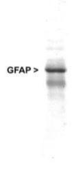
- Western Blot: GFAP Antibody (GTX27260) - Western blot analysis of GFAP expression in whole rat cerebellum homogenate using GTX27260

Supportive validation
- Submitted by
- GeneTex (provider)
- Main image
- Experimental details
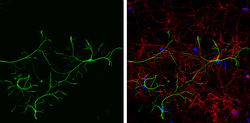
- GFAP antibody detects GFAP protein expression in astrocytes and Tuj1 antibody detects Tuj1 protein expression in neurons by immunofluorescent analysis.Sample:Cultured rat E18 primary cortical neurons and astrocytes, DIV 8. Cells were fixed in 4% paraformaldehyde at RT for 15 min.Green: GFAP protein stained by GFAP antibody (GTX27260) diluted at 1:250.Red: beta Tubulin 3/ TUJ1, stained by beta Tubulin 3/ TUJ1 antibody [GT11710] (GTX631836) diluted at 1:250.Blue: Fluoroshield with DAPI (GTX30920).

- Submitted by
- GeneTex (provider)
- Main image
- Experimental details
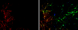
- GFAP antibody detects GFAP protein expression by immunofluorescent analysis.Sample:Cultured rat E18 primary cortical neuron, DIV 8 cells were fixed in 4% paraformaldehyde at RT for 15 min.Red: GFAP protein stained by GFAP antibody (GTX27260) diluted at 1:250.Green: Glutamine synthetase, stained by Glutamine synthetase antibody [GT7711] (GTX30657) diluted at 1:250.Blue: Fluoroshield with DAPI (GTX30920).

- Submitted by
- GeneTex (provider)
- Main image
- Experimental details
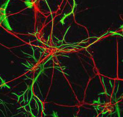
- Immunocytochemistry/Immunofluorescence: GFAP Antibody (GTX27260) - Rat neurons stained with Neurofilament Heavy antibody (red) and GFAP antibody GTX27260 (green).

- Submitted by
- GeneTex (provider)
- Main image
- Experimental details
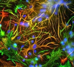
- Immunocytochemistry/Immunofluorescence: GFAP Antibody (GTX27260) - Mixed neuron-glial cultures stained with GFAP antibody GTX27260 (red) and Vimentin antibody GTX30668 (green). The fibroblastic cells contain only Vimentin and so are green. The astrocytes contain either Vimentin and GFAP (appearing golden) or predominantly GFAP (appearing red). Blue is nuclear DNA stain.
