Explore
 Validate
 Learn
Learn Western blot
 Flow cytometry
Antibody data
- Antibody Data
- Antigen structure
- References [2]
- Comments [0]
- Validations
- Western blot [1]
- Immunohistochemistry [1]
- Other assay [1]
Submit
Validation data
Reference
Comment
Report error
- Product number
- MA5-16383 - Provider product page

- Provider
- Invitrogen Antibodies
- Product name
- Myeloperoxidase Monoclonal Antibody (SP72)
- Antibody type
- Monoclonal
- Antigen
- Synthetic peptide
- Description
- Heat-mediated antigen retrieval is recommended prior to staining, using a 10mM citrate buffer, pH 6.0, for 10 minutes followed by cooling at room temperature for 20 min. Following antigen retrieval, incubate samples with primary antibody for 30 min at room temperature. A suggested positive control is tonsil tissue.
- Reactivity
- Human
- Host
- Rabbit
- Isotype
- IgG
- Antibody clone number
- SP72
- Vial size
- 500 µL
- Concentration
- 0.212 mg/mL
- Storage
- 4° C
Submitted references Synthetic Siglec-9 Agonists Inhibit Neutrophil Activation Associated with COVID-19.
Potential roles of interleukin-17A in the development of skin fibrosis in mice.
Delaveris CS, Wilk AJ, Riley NM, Stark JC, Yang SS, Rogers AJ, Ranganath T, Nadeau KC, Stanford COVID-19 Biobank, Blish CA, Bertozzi CR
ChemRxiv : the preprint server for chemistry 2020 Dec 17;
ChemRxiv : the preprint server for chemistry 2020 Dec 17;
Potential roles of interleukin-17A in the development of skin fibrosis in mice.
Okamoto Y, Hasegawa M, Matsushita T, Hamaguchi Y, Huu DL, Iwakura Y, Fujimoto M, Takehara K
Arthritis and rheumatism 2012 Nov;64(11):3726-35
Arthritis and rheumatism 2012 Nov;64(11):3726-35
No comments: Submit comment
Supportive validation
- Submitted by
- Invitrogen Antibodies (provider)
- Main image
- Experimental details
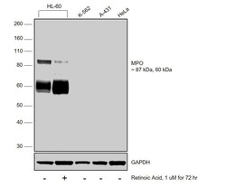
- Western blot was performed using Anti-Myeloperoxidase Monoclonal Antibody (SP72) (Product # MA5-16383) and 87 kDa, 60 kDa bands corresponding to MPO was observed across cell lines except K-562, A-431 and HeLa and was observed to decrease upon Retinoic Acid treatment. Membrane enriched extracts (30 µg lysate) of HL-60 (Lane 1), HL-60 treated with Retinoic Acid (1uM for 72 Hours) (Lane 2), K-562 (Lane 3), A-431 (Lane 4) and HeLa (Lane 5) were electrophoresed using NuPAGE™ 4-12% Bis-Tris Protein Gel (Product # NP0322BOX). Resolved proteins were then transferred onto a nitrocellulose membrane (Product # IB23001) by iBlot® 2 Dry Blotting System (Product # IB21001). The blot was probed with the primary antibody (1:1000 dilution) and detected by chemiluminescence with Goat anti-Mouse IgG (H+L), Superclonal™ Recombinant Secondary Antibody, HRP (Product # A28177, 1:4000 dilution) using the iBright FL 1000 (Product # A32752). Chemiluminescent detection was performed using Novex® ECL Chemiluminescent Substrate Reagent Kit (Product # WP20005).

Supportive validation
- Submitted by
- Invitrogen Antibodies (provider)
- Main image

- Experimental details
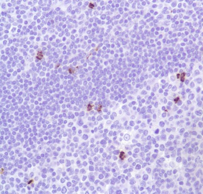
- Immunohistochemical analysis of Myeloperoxidase using anti-Myeloperoxidase Monoclonal Antibody (Product # MA5-16383) in Tonsil Tissue. The recommened dilution for this antibody in immunohistochemistry applications is 1:100.
Supportive validation
- Submitted by
- Invitrogen Antibodies (provider)
- Main image

- Experimental details
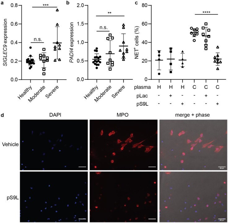
- Figure 4. A Siglec-9 agonist inhibits NETosis of neutrophils induced by COVID-19 plasma. ( a , b ) Analysis of publicly available single-cell transcriptomics data for SIGLEC9 expression ( a ) and PADI4 expression ( b ) on neutrophils in peripheral blood from healthy donors or COVID-19 patients. Error bars represent SD. Statistics were determined using mixed effects model. ** = p < 0.01; *** = p < 0.001 ( c,d ) Primary neutrophils were cultured in undiluted and citrate anticoagulated plasma from healthy donors or COVID-19 patients for 4 h. Cells were fixed, stained for extracellular myeloperoxidase, and imaged in DAPI imaging media by fluorescence microscopy. Cells were treated in technical triplicate and imaged across multiple fields of view. ( c ) Proportion of NET-positive cells (%) across all fields of view. Each dot represents and individual plasma sample. ( d ) Representative images from a COVID-19 patient plasma sample with or without pS9L. Error bars represent SD. Statistics were determined using mixed effects models to account for samples using repeat neutrophil donors. **** = p < 0.0001.