 Explore
 Validate
 Learn
Learn Immunohistochemistry
Antibody data
- Antibody Data
- Antigen structure
- References [8]
- Comments [0]
- Validations
- Immunohistochemistry [1]
- Other assay [2]
Submit
Validation data
Reference
Comment
Report error
- Product number
- 14-9751-82 - Provider product page

- Provider
- Invitrogen Antibodies
- Product name
- Somatostatin Monoclonal Antibody (ICDCLS), eBioscience™
- Antibody type
- Monoclonal
- Antigen
- Other
- Description
- Description: The monoclonal antibody ICDCLS recognizes human, mouse, and rat Somatostatin. Somatostatin, also known as growth hormone-inhibiting hormone (GHIH) or somatotrophin release-inhibiting factor (SRIF), is a peptide hormone with two active forms (14 and 28 amino acids) produced by alternative cleavage of a single preproprotein. Somatostatin is secreted by the delta cells of the islet of Langerhans in the pancreas as well as by the hypothalamus and intestine. Somatostatin has a variety of functions, such as regulating the endocrine system and affecting neurotransmission and cell proliferation. Somatostatin also inhibits the release of numerous secondary hormones, such as insulin and glucagon. Somatostatin can be used to identify medium size aspiny neurons in the striatum, cortex, and hippocampus. Expression of somatostatin is present in hyperplasia of the pancreatic islets as well as islet cell tumors such as somatostatinomas.
- Antibody clone number
- ICDCLS
- Concentration
- 0.5 mg/mL
Submitted references Ciliary neuropeptidergic signaling dynamically regulates excitatory synapses in postnatal neocortical pyramidal neurons.
PDX1(LOW) MAFA(LOW) β-cells contribute to islet function and insulin release.
Vitamin-D-Binding Protein Contributes to the Maintenance of α Cell Function and Glucagon Secretion.
Super-resolution microscopy compatible fluorescent probes reveal endogenous glucagon-like peptide-1 receptor distribution and dynamics.
Somatostatin and somatostatin receptors.
Substance P receptor-immunoreactive neurons in the rat neostriatum are segregated into somatostatinergic and cholinergic aspiny neurons.
"Somatostatinoma": a somatostatin-containing tumor of the endocrine pancreas.
Hypertrophy and hyperplasia of somatostatin-containing D-cells in diabetes.
Tereshko L, Gao Y, Cary BA, Turrigiano GG, Sengupta P
eLife 2021 Mar 2;10
eLife 2021 Mar 2;10
PDX1(LOW) MAFA(LOW) β-cells contribute to islet function and insulin release.
Nasteska D, Fine NHF, Ashford FB, Cuozzo F, Viloria K, Smith G, Dahir A, Dawson PWJ, Lai YC, Bastidas-Ponce A, Bakhti M, Rutter GA, Fiancette R, Nano R, Piemonti L, Lickert H, Zhou Q, Akerman I, Hodson DJ
Nature communications 2021 Jan 29;12(1):674
Nature communications 2021 Jan 29;12(1):674
Vitamin-D-Binding Protein Contributes to the Maintenance of α Cell Function and Glucagon Secretion.
Viloria K, Nasteska D, Briant LJB, Heising S, Larner DP, Fine NHF, Ashford FB, da Silva Xavier G, Ramos MJ, Hasib A, Cuozzo F, Manning Fox JE, MacDonald PE, Akerman I, Lavery GG, Flaxman C, Morgan NG, Richardson SJ, Hewison M, Hodson DJ
Cell reports 2020 Jun 16;31(11):107761
Cell reports 2020 Jun 16;31(11):107761
Super-resolution microscopy compatible fluorescent probes reveal endogenous glucagon-like peptide-1 receptor distribution and dynamics.
Ast J, Arvaniti A, Fine NHF, Nasteska D, Ashford FB, Stamataki Z, Koszegi Z, Bacon A, Jones BJ, Lucey MA, Sasaki S, Brierley DI, Hastoy B, Tomas A, D'Agostino G, Reimann F, Lynn FC, Reissaus CA, Linnemann AK, D'Este E, Calebiro D, Trapp S, Johnsson K, Podewin T, Broichhagen J, Hodson DJ
Nature communications 2020 Jan 24;11(1):467
Nature communications 2020 Jan 24;11(1):467
Somatostatin and somatostatin receptors.
Kumar U, Grant M
Results and problems in cell differentiation 2010;50:137-84
Results and problems in cell differentiation 2010;50:137-84
Substance P receptor-immunoreactive neurons in the rat neostriatum are segregated into somatostatinergic and cholinergic aspiny neurons.
Kaneko T, Shigemoto R, Nakanishi S, Mizuno N
Brain research 1993 Dec 24;631(2):297-303
Brain research 1993 Dec 24;631(2):297-303
"Somatostatinoma": a somatostatin-containing tumor of the endocrine pancreas.
Ganda OP, Weir GC, Soeldner JS, Legg MA, Chick WL, Patel YC, Ebeid AM, Gabbay KH, Reichlin S
The New England journal of medicine 1977 Apr 28;296(17):963-7
The New England journal of medicine 1977 Apr 28;296(17):963-7
Hypertrophy and hyperplasia of somatostatin-containing D-cells in diabetes.
Orci L, Baetens D, Rufener C, Amherdt M, Ravazzola M, Studer P, Malaisse-Lagae F, Unger RH
Proceedings of the National Academy of Sciences of the United States of America 1976 Apr;73(4):1338-42
Proceedings of the National Academy of Sciences of the United States of America 1976 Apr;73(4):1338-42
No comments: Submit comment
Supportive validation
- Submitted by
- Invitrogen Antibodies (provider)
- Main image

- Experimental details
- Immunohistochemistry of formalin-fixed paraffin embedded human pancreas tissue using 0.1 µg/mL of Mouse IgG1 K Isotype Control Purified (left) or 0.1 µg/mL of Anti-Somatostatin Purified (right) followed by Anti-Mouse IgG Biotin, Streptavidin-HRP, and DAB visualization.Nuclei are counterstained with hematoxylin.

Supportive validation
- Submitted by
- Invitrogen Antibodies (provider)
- Main image
- Experimental details
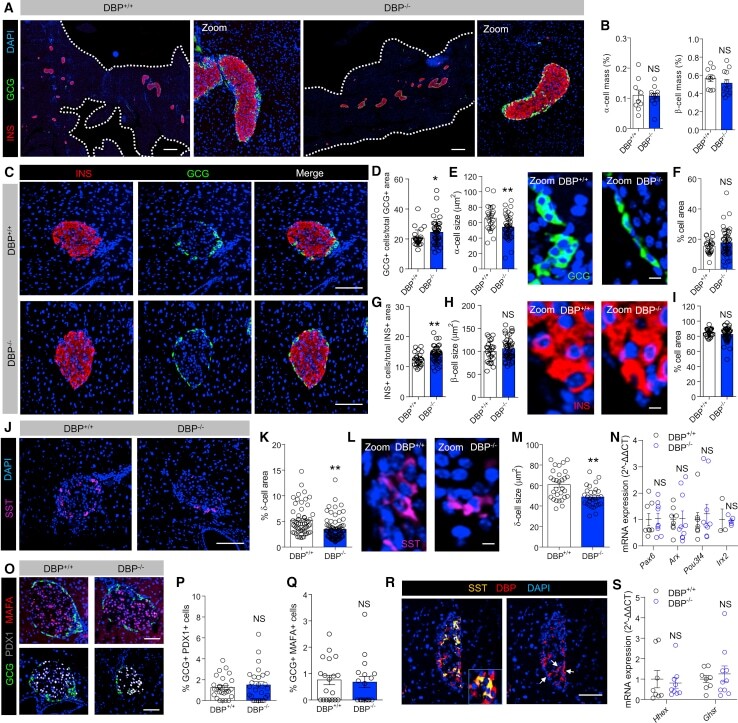
- Figure 2 DBP Alters alpha Cell and delta Cell Number and Size (A and B) Cell resolution reconstruction of pancreatic sections reveals no differences in alpha cell and beta cell mass in DBP +/+ and DBP -/- mice (A), quantified in bar graph (B) (scale bar represents 530 mum; representative images are shown; inset is a zoom showing maintenance of cellular resolution in a single image; n = 9-12 sections from 3 to 4 animals; unpaired t test). (C-I) Morphological analyses of DBP -/- islets (C) reveal increased alpha cell number (D) and decreased alpha cell size (E) (representative images shown in right panel), but normal area occupied (F). By contrast, beta cell number is increased (G), although beta cell size (H) (representative images shown in right panel) and area (I) are unchanged (scale bar in D represents 85 mum; scale bars in E and H represent 10 mum; n = 24-45 islets from 3 to 4 animals; E and H are zooms of C to better show alpha cell and beta cell size; DAPI is shown in blue; unpaired t test). (J-M) delta cell proportion (J and K; n = 55-79 islets from 4 to 5 animals) and size (L and M; n = 29 to 30 islets from 3 animals) are decreased in DBP -/- islets (scale bar in J represents 85 mum; scale bar in L represents 10 mum; representative images are shown; L is a zoom of J to better show delta cell size; unpaired t test). (N) Expression levels of the alpha cell differentiation markers Arx , Pax6 , Pou3f4 , and Irx2 are similar in DBP +/- and DBP -/- islets (n = 3-10 animals;
- Submitted by
- Invitrogen Antibodies (provider)
- Main image

- Experimental details
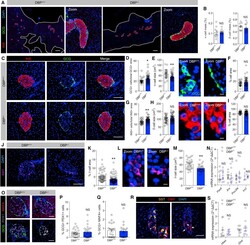
- Fig. 4 LUXendin645 reveals GLP1R expression in a subpopulation of alpha-cells. a - c LUXendin645 labeling is widespread throughout the intact islet, co-localizing predominantly with beta-cells a and delta-cells b , but less so with alpha-cells c stained for insulin (INS), somatostatin (SST), and glucagon (GCG), respectively ( n = 18 islets, seven animals, three separate islet preparations) (scale bar = 26 um). d Following dissociation of islets into cell clusters, LUXendin645 labeling can be more accurately quantified (arrows highlight cells selected for zoom-in) (scale bar = 26 um). e Zoom-in of d showing a LUXendin645 - (left) and LUXendin645 + (right) alpha-cell (arrows highlight non-labeled cell membrane, which is not bounded by a beta-cell) (scale bar = 26 um). f Box-and-whiskers plot showing proportion of beta-cells (INS) and alpha-cells (GCG) co-localized with LUXendin645 ( n = 18 cell clusters, ten animals, three separate islet preparations) (box and whiskers plot shows range and median; mean is shown by a plus symbol). g Ins1Cre Thor ;R26 mT/mG dual fluorophore reporter islets express tdTomato until Cre-mediated replacement with mGFP, allowing identification of beta-cells (~80% of the islet population) and non-beta-cells for live imaging (scale bar = 26 um). LUXendin645 (LUX645) highlights GLP1R expression in nearly all beta-cells but relatively few non-beta-cells ( n = 31 islets, six animals, three separate islet preparations). h A zoom
