Explore
 Validate
 Learn
Learn Western blot
Antibody data
- Antibody Data
- Antigen structure
- References [0]
- Comments [0]
- Validations
- Western blot [4]
- Immunocytochemistry [1]
- Immunohistochemistry [2]
- Flow cytometry [1]
Submit
Validation data
Reference
Comment
Report error
- Product number
- MA5-17075 - Provider product page

- Provider
- Invitrogen Antibodies
- Product name
- Fibronectin Monoclonal Antibody (2F4)
- Antibody type
- Monoclonal
- Antigen
- Purifed from natural sources
- Description
- MA5-17075 targets FN1 in FACS, IHC and WB applications and shows reactivity with Human samples.
- Antibody clone number
- 2F4
- Concentration
- Conc. Not Determined
No comments: Submit comment
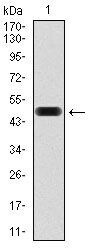
Supportive validation
- Submitted by
- Invitrogen Antibodies (provider)
- Main image

- Experimental details
- Western blot analysis of FN1 using a FN1 monoclonal antibody (Product # MA5-17075) against a human FN1 (AA: 1965-2176) recombinant protein.

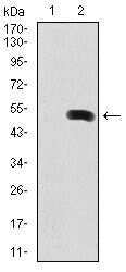
- Submitted by
- Invitrogen Antibodies (provider)
- Main image

- Experimental details
- Western blot analysis of FN1 using FN1 monoclonal antibody (Product # MA5-17075) in HEK293 (1) and FN1 (AA: 1965-2176) human IgG Fc transfected HEK293 (2) cell lysate.
- Submitted by
- Invitrogen Antibodies (provider)
- Main image

- Experimental details
- Western blot analysis of FN1 using a FN1 monoclonal antibody (Product # MA5-17075) against a human FN1 (AA: 1965-2176) recombinant protein.
- Submitted by
- Invitrogen Antibodies (provider)
- Main image
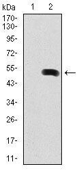
- Experimental details
- Western blot analysis of FN1 using FN1 monoclonal antibody (Product # MA5-17075) in HEK293 (1) and FN1 (AA: 1965-2176) human IgG Fc transfected HEK293 (2) cell lysate.

Supportive validation
- Submitted by
- Invitrogen Antibodies (provider)
- Main image
- Experimental details
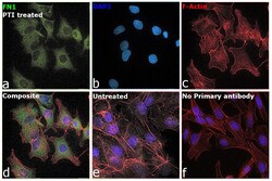
- Immunofluorescence analysis of Fibronectin was performed using 70% confluent log phase Hep G2 cells and in HepG2 cells treated with PTI. The cells were fixed with 4% paraformaldehyde for 10 minutes, permeabilized with 0.1% Triton™ X-100 for 15 minutes, and blocked with 2% BSA for 45 minutes at room temperature. The cells were labeled with Fibronectin Monoclonal Antibody (2F4) (Product # MA5-17075) at 1:100 in 0.1% BSA, incubated at 4 degree celsius overnight and then labeled with Donkey anti-Mouse IgG (H+L) Highly Cross-Adsorbed Secondary Antibody, Alexa Fluor Plus 488 (Product # A32766), (1:2000), for 45 minutes at room temperature (Panel a: Green). Nuclei (Panel b:Blue) were stained with ProLong™ Diamond Antifade Mountant with DAPI (Product # P36962). F-actin (Panel c: Red) was stained with Rhodamine Phalloidin (Product # R415, 1:300). Panel d represents the merged image showing Plasma membrane and cytoplasm localization. Panel e represents untreated HepG2 cells and Panel f represents control cells with no primary antibody to assess background. The images were captured at 60X magnification.

Supportive validation
- Submitted by
- Invitrogen Antibodies (provider)
- Main image
- Experimental details
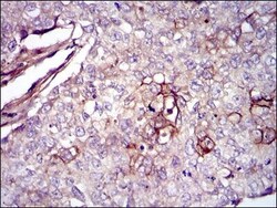
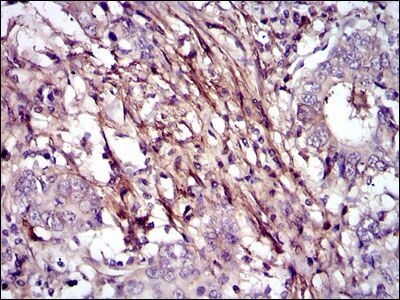
- Immunohistochemical analysis of paraffin-embedded lung cancer tissues using FN1 monoclonal antibody (Product # MA5-17075) followed with DAB staining.
- Submitted by
- Invitrogen Antibodies (provider)
- Main image

- Experimental details
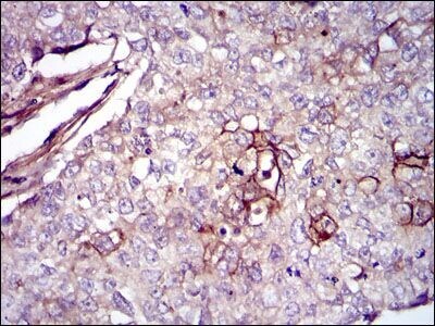
- Immunohistochemical analysis of paraffin-embedded stomach cancer tissues using FN1 monoclonal antibody (Product # MA5-17075) followed with DAB staining.
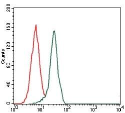
Supportive validation
- Submitted by
- Invitrogen Antibodies (provider)
- Main image

- Experimental details
- Flow cytometric analysis of HeLa cells using FN1 monoclonal antibody (Product # MA5-17075) (green) and negative control (red).