Explore
 Validate
 Learn
Learn Immunocytochemistry
Antibody data
- Antibody Data
- Antigen structure
- References [8]
- Comments [0]
- Validations
- Immunocytochemistry [1]
- Chromatin Immunoprecipitation [1]
Submit
Validation data
Reference
Comment
Report error
- Product number
- MA1-7629 - Provider product page

- Provider
- Invitrogen Antibodies
- Product name
- p53 Monoclonal Antibody (BD53-12)
- Antibody type
- Monoclonal
- Antigen
- Other
- Description
- MA1-7629 detects p53 from human, rat and mouse samples.
- Antibody clone number
- BD53-12
- Concentration
- 1 mg/mL
Submitted references The effect of vitamin C administration on monosodium glutamate induced liver injury. An experimental study.
The effects of PDE5 inhibitory drugs on renal ischemia/reperfusion injury in rats.
Hepatoprotective effect of coenzyme Q10 in rats with acetaminophen toxicity.
X-irradiation reduces the proliferation of astrocytes by cell cycle arrest.
Chk1 deficiency in the mouse small intestine results in p53-independent crypt death and subsequent intestinal compensation.
Protective effects of doxycycline in ischemia/reperfusion injury on kidney.
Immunohistochemical study of proteins linked to apoptosis in rat fetal kidney cells following prepregnancy adriamycin administration in the mother.
p53 Expression and apoptosis in liver and spleen during CO2 pneumoperitoneum.
El-Meghawry El-Kenawy A, Osman HE, Daghestani MH
Experimental and toxicologic pathology : official journal of the Gesellschaft fur Toxikologische Pathologie 2013 Jul;65(5):513-21
Experimental and toxicologic pathology : official journal of the Gesellschaft fur Toxikologische Pathologie 2013 Jul;65(5):513-21
The effects of PDE5 inhibitory drugs on renal ischemia/reperfusion injury in rats.
Küçük A, Yucel M, Erkasap N, Tosun M, Koken T, Ozkurt M, Erkasap S
Molecular biology reports 2012 Oct;39(10):9775-82
Molecular biology reports 2012 Oct;39(10):9775-82
Hepatoprotective effect of coenzyme Q10 in rats with acetaminophen toxicity.
Fouad AA, Jresat I
Environmental toxicology and pharmacology 2012 Mar;33(2):158-67
Environmental toxicology and pharmacology 2012 Mar;33(2):158-67
X-irradiation reduces the proliferation of astrocytes by cell cycle arrest.
Wang Q, Xu Y, Xie MJ, Yu ZY, Qin YY, Wang W, Zhu Z
Neuroscience letters 2011 Jul 1;498(1):78-83
Neuroscience letters 2011 Jul 1;498(1):78-83
Chk1 deficiency in the mouse small intestine results in p53-independent crypt death and subsequent intestinal compensation.
Greenow KR, Clarke AR, Jones RH
Oncogene 2009 Mar 19;28(11):1443-53
Oncogene 2009 Mar 19;28(11):1443-53
Protective effects of doxycycline in ischemia/reperfusion injury on kidney.
Kucuk A, Kabadere S, Tosun M, Koken T, Kinaci MK, Isikli B, Erkasap N
Journal of physiology and biochemistry 2009 Jun;65(2):183-91
Journal of physiology and biochemistry 2009 Jun;65(2):183-91
Immunohistochemical study of proteins linked to apoptosis in rat fetal kidney cells following prepregnancy adriamycin administration in the mother.
Pedrycz A, Czerny K
Acta histochemica 2008;110(6):519-23
Acta histochemica 2008;110(6):519-23
p53 Expression and apoptosis in liver and spleen during CO2 pneumoperitoneum.
Arikan Y, Tosun M, Saykol V, Kalkan S, Erdem S
Langenbeck's archives of surgery 2008 Nov;393(6):877-82
Langenbeck's archives of surgery 2008 Nov;393(6):877-82
No comments: Submit comment
Supportive validation
- Submitted by
- Invitrogen Antibodies (provider)
- Main image

- Experimental details
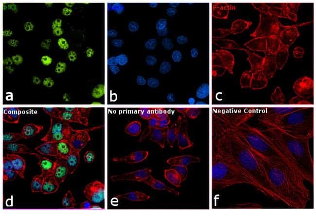
- Immunofluorescence analysis of p53 was performed using 70% confluent log phaseT-47D cells. The cells were fixed with 4% paraformaldehyde for 10 minutes, permeabilized with 0.1% Triton™ X-100 for 10 minutes, and blocked with 1% BSA for 1 hour at room temperature. The cells were labeled with p53 Monoclonal Antibody (4A8) (Product # MA1-7629) at 5 µg/mL in 0.1% BSA and incubated overnight at 4 degree Celsius and then labeled with Goat anti-Mouse IgG (H+L) Superclonal™ Secondary Antibody, Alexa Fluor® 488 conjugate (Product # A28175) at a dilution of 1:2000 for 45 minutes at room temperature (Panel a: green). Nuclei (Panel b: blue) were stained with SlowFade® Gold Antifade Mountant with DAPI (Product # S36938). F-actin (Panel c: red) was stained with Rhodamine Phalloidin (Product # R415, 1:300). Panel d represents the merged image showing nuclear localization. Panel f represents SK-OV-3 cells as negative control, showing no p53 staining. Panel e represents control cells with no primary antibody to assess background. The images were captured at 60X magnification.
Supportive validation
- Submitted by
- Invitrogen Antibodies (provider)
- Main image

- Experimental details
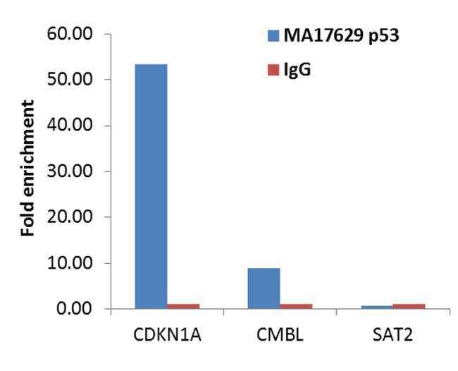
- Enrichment of endogenous p53 protein at specific gene loci using Anti-p53 Antibody: Chromatin Immunoprecipitation (ChIP) was performed using Anti-p53 Mouse Monoclonal Antibody (Product # MA1-7629, 4 µg) on sheared chromatin from 2 million Etoposide-treated A-431 cells using the MAGnify ChIP system kit (Product # 49-2024). Normal Rabbit IgG was used as a negative IP control. The purified DNA was analyzed by qPCR with PCR primer pairs over the CDKN1A and CMBL genes (active) and SAT2 satellite repeats (inactive). Data is presented as fold enrichment of the antibody signal versus the negative control IgG using the comparative CT method.