Explore
 Validate
 Learn
Learn Western blot
Antibody data
- Antibody Data
- Antigen structure
- References [3]
- Comments [0]
- Validations
- Western blot [1]
- Immunocytochemistry [2]
- Immunoprecipitation [1]
- Immunohistochemistry [1]
Submit
Validation data
Reference
Comment
Report error
- Product number
- GTX100629 - Provider product page

- Provider
- GeneTex
- Proper citation
- GeneTex Cat#GTX100629, RRID:AB_2037587
- Product name
- p53 antibody [N1], N-term
- Antibody type
- Polyclonal
- Reactivity
- Human
- Host
- Rabbit
Submitted references DNA damage induced by topoisomerase inhibitors activates SAMHD1 and blocks HIV-1 infection of macrophages.
Stimulation of Toll-like receptor-1/2 combined with Velcade increases cytotoxicity to human multiple myeloma cells.
The synthetic flavonoid WYC02-9 inhibits colorectal cancer cell growth through ROS-mediated activation of MAPK14 pathway.
Mlcochova P, Caswell SJ, Taylor IA, Towers GJ, Gupta RK
The EMBO journal 2018 Jan 4;37(1):50-62
The EMBO journal 2018 Jan 4;37(1):50-62
Stimulation of Toll-like receptor-1/2 combined with Velcade increases cytotoxicity to human multiple myeloma cells.
Abdi J, Mutis T, Garssen J, Redegeld F
Blood cancer journal 2013 May 31;3:e119
Blood cancer journal 2013 May 31;3:e119
The synthetic flavonoid WYC02-9 inhibits colorectal cancer cell growth through ROS-mediated activation of MAPK14 pathway.
Chen YJ, Chen HP, Cheng YJ, Lin YH, Liu KW, Chen YJ, Hou MF, Wu YC, Lee YC, Yuan SS
Life sciences 2013 Jun 13;92(22):1081-92
Life sciences 2013 Jun 13;92(22):1081-92
No comments: Submit comment
Supportive validation
- Submitted by
- GeneTex (provider)
- Main image
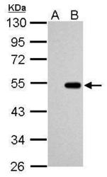
- Experimental details
- Sample (30 ug of whole cell lysate) A: HCT116 cells with mock treatment for 24hr B: HCT116 cells with 30uM cisplatin treatment for 24hr 10% SDS PAGE GTX100629 diluted at 1:1000
Supportive validation
- Submitted by
- GeneTex (provider)
- Main image

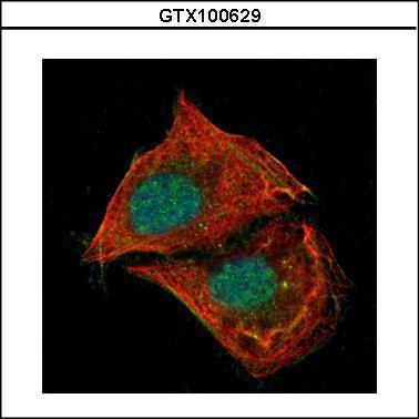
- Experimental details
- Confocal immunofluorescence analysis (Olympus FV10i) of paraformaldehyde-fixed U2OS, using p53(GTX100629) antibody (Green) at 1:500 dilution. Alpha-tubulin filaments were labeled with GTX11304 (Red) at 1:2000.
- Submitted by
- GeneTex (provider)
- Main image
- Experimental details
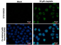
- p53 antibody [N1], N-term detects p53 protein at nucleus by immunofluorescent analysis. Samples: HCT 116 cells mock (left) and treated with 30 £gM Cisplatin for 24 hrs (right) were fixed in 4% paraformaldehyde at RT for 15 min.Green: p53 protein stained by p53 antibody [N1], N-term (GTX100629) diluted at 1:500.Blue: Hoechst 33342 staining.Scale bar = 10 £gm.

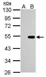
Supportive validation
- Submitted by
- GeneTex (provider)
- Main image
- Experimental details
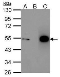
- Immunoprecipitation of p53 protein. HCT116 lysates with 30uM cisplatin treatment for 24 hours were subjected to immunoprecipitation using (B) normal rabbit IgG or (C) 2.5 ug of anti-p53 antibody (GTX100629). (A) Input, 20ug of HCT116 lysates. The precipitated protein was detected by GTX100629 diluted at 1:10000. EasyBlot anti-Rabbit IgG Kit (GTX225856-01) was used in Western blot.

Supportive validation
- Submitted by
- GeneTex (provider)
- Main image
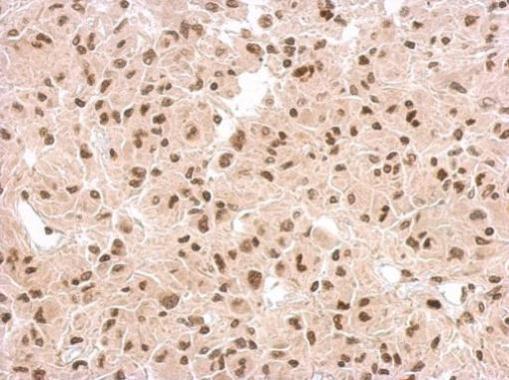
- Experimental details
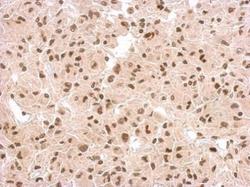
- Immunohistochemical analysis of paraffin-embedded U373 xenograft, using p53(GTX100629) antibody at 1:500 dilution.