Explore
 Validate
 Learn
Learn Western blot
 ELISA
ELISAAntibody data
- Antibody Data
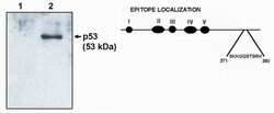
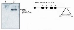
- Antigen structure
- References [0]
- Comments [0]
- Validations
- Western blot [3]
- Immunocytochemistry [1]
Submit
Validation data
Reference
Comment
Report error
- Product number
- MA1-12648 - Provider product page

- Provider
- Invitrogen Antibodies
- Product name
- p53 Monoclonal Antibody (HR231)
- Antibody type
- Monoclonal
- Antigen
- Recombinant full-length protein
- Description
- A suggested positive control for this product is breast or colon carcinoma cells.
- Reactivity
- Human, Mouse, Rat, Simian
- Host
- Mouse
- Isotype
- IgG
- Antibody clone number
- HR231
- Vial size
- 100 µg
- Concentration
- 1 mg/mL
- Storage
- -20° C, Avoid Freeze/Thaw Cycles
No comments: Submit comment
Supportive validation
- Submitted by
- Invitrogen Antibodies (provider)
- Main image
- Experimental details
- Western blot analysis of p53 using a monoclonal antibody (Product # MA1-12648).

- Submitted by
- Invitrogen Antibodies (provider)
- Main image
- Experimental details
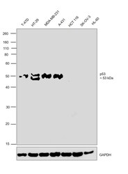
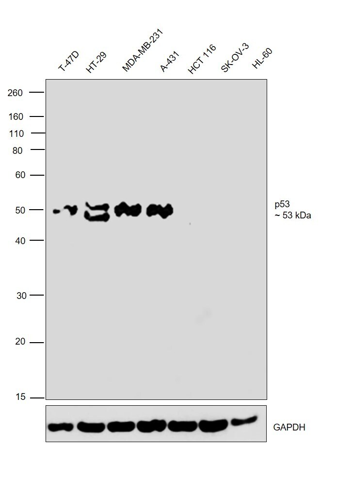
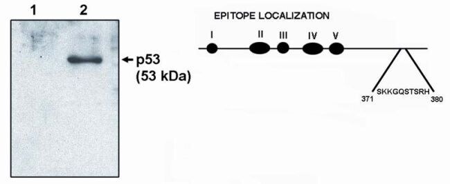
- Western blot was performed using Anti-p53 Monoclonal Antibody (HR231) (Product # MA1-12648) and a 53 kDa band corresponding to Cellular tumor antigen p53 was observed across cell lines except HCT 116, SK-OV-3 and HL-60 which are reported to be negative. Modified whole cell extracts (1%SDS) (30 µg lysate) of T-47D (Lane 1), HT-29 (Lane 2), MDA-MB-231 (Lane 3), A-431 (Lane 4), HCT 116 (Lane 5), SK-O-V3 (Lane 6) and HL-60 (Lane 7) were electrophoresed using NuPAGE™ 10% Bis-Tris Protein Gel (Product # NP0302BOX). Resolved proteins were then transferred onto a Nitrocellulose membrane (Product # IB23001) by iBlot® 2 Dry Blotting System (Product # IB21001). The blot was probed with the primary antibody (2 µg/mL) and detected by chemiluminescence with Goat anti-Mouse IgG (H+L) Superclonal™ Recombinant Secondary Antibody, HRP (Product # A28177, 1:4000 dilution) using the iBright FL 1000 (Product # A32752). Chemiluminescent detection was performed using Novex® ECL Chemiluminescent Substrate Reagent Kit (Product # WP20005).
- Submitted by
- Invitrogen Antibodies (provider)
- Main image
- Experimental details
- Western blot analysis of p53 using a monoclonal antibody (Product # MA1-12648).
Supportive validation
- Submitted by
- Invitrogen Antibodies (provider)
- Main image
- Experimental details
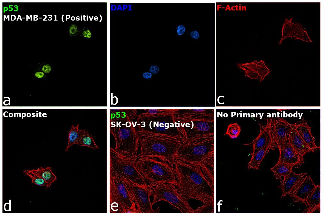
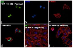
- Immunofluorescence analysis of Cellular tumor antigen p53 was performed using 70% confluent log phase MDA-MB-231 cells. The cells were fixed with 4% paraformaldehyde for 10 minutes, permeabilized with 0.1% Triton™ X-100 for 15 minutes, and blocked with 2% BSA for 45 minutes at room temperature. The cells were labeled with p53 Monoclonal Antibody (HR231) (Product # MA1-12648) at 1:100 dilution in 0.1% BSA, incubated at 4 degree celsius overnight and then labeled with Donkey anti-Mouse IgG (H+L) Highly Cross-Adsorbed Secondary Antibody, Alexa Fluor Plus 488 (Product # A32766), (1:2000 dilution), for 45 minutes at room temperature (Panel a: Green). Nuclei (Panel b: Blue) were stained with ProLong™ Diamond Antifade Mountant with DAPI (Product # P36962). F-actin (Panel c: Red) was stained with Rhodamine Phalloidin (Product # R415, 1:300). Panel d represents the merged image showing Nuclear localization. Panel e represents SK-OV-3 cells having no expression of p53. Panel f represents control cells with no primary antibody to assess background. The images were captured at 60X magnification.