 Explore
 Validate
 Learn
LearnPA5-48092
antibody from Invitrogen Antibodies
Targeting: AIFM1
AIF, AUNX1, CMTX4, DFNX5, NAMSD, PDCD8
 Western blot
Antibody data
- Antibody Data
- Antigen structure
- References [0]
- Comments [0]
- Validations
- Western blot [4]
- Immunohistochemistry [1]
Submit
Validation data
Reference
Comment
Report error
- Product number
- PA5-48092 - Provider product page

- Provider
- Invitrogen Antibodies
- Product name
- AIF Polyclonal Antibody
- Antibody type
- Polyclonal
- Antigen
- Recombinant full-length protein
- Description
- Reconstitute at 0.2 mg/mL in sterile PBS.
- Reactivity
- Human, Mouse, Rat
- Host
- Sheep
- Isotype
- IgG
- Vial size
- 100 µg
- Concentration
- 0.2 mg/mL
- Storage
- -20° C, Avoid Freeze/Thaw Cycles
No comments: Submit comment
Supportive validation
- Submitted by
- Invitrogen Antibodies (provider)
- Main image
- Experimental details
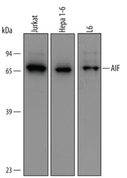
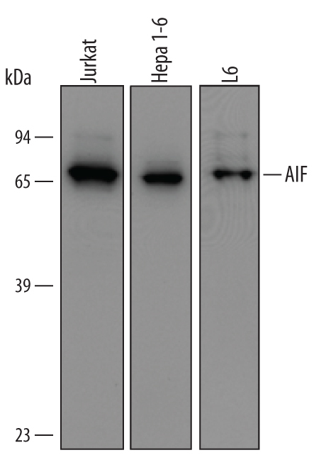
- Western blot analysis from lysates of Jurkat human acute T cell leukemia cell line, Hepa 1-6 mouse hepatoma cell line, and L6 rat myoblast cell line. PVDF membrane was probed with 1 µg/mL of Sheep Anti-human/mouse/Rat AIF Antigen Affinity-purified Polyclonal Antibody (Product # PA5-48092) followed by HRP-conjugated Anti-Sheep IgG Secondary Antibody. A specific band was detected for AIF at approximately 65 kDa (as indicated). This experiment was conducted under reducing conditions.
- Submitted by
- Invitrogen Antibodies (provider)
- Main image
- Experimental details
- Western blot analysis of AIF in Jurkat human acute T cell leukemia cell line, Hepa 1‚6 mouse hepatoma cell line, and L6 rat myoblast cell line. Samples were incubated in AIF polyclonal antibody (Product # PA5-48092) using a dilution of 1 µg/mL followed by a HRP-conjugated Anti-Sheep IgG secondary antibody. A specific band was detected for AIF at approximately 65 kDa (as indicated). This experiment was conducted under reducing conditions.
- Submitted by
- Invitrogen Antibodies (provider)
- Main image
- Experimental details
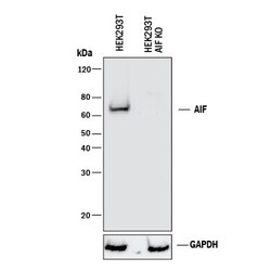
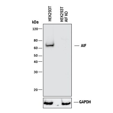
- Knockout validation by Western blot analysis of AIF in lysates of HEK293T human embryonic kidney parental cell line and AIF knockout HEK293T cell line (KO). Samples were incubated in AIF polyclonal antibody (Product # PA5-48092) using a dilution of 1 µg/mL followed by a HRP-conjugated Anti-Sheep IgG secondary antibody. A specific band was detected for AIF at approximately 65 kDa (as indicated) in the parental HEK293T cell line, but is not detectable in knockout HEK293T cell line. GAPDH is shown as a loading control. This experiment was conducted under reducing conditions.
- Submitted by
- Invitrogen Antibodies (provider)
- Main image

- Experimental details
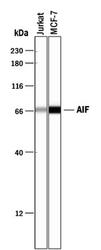
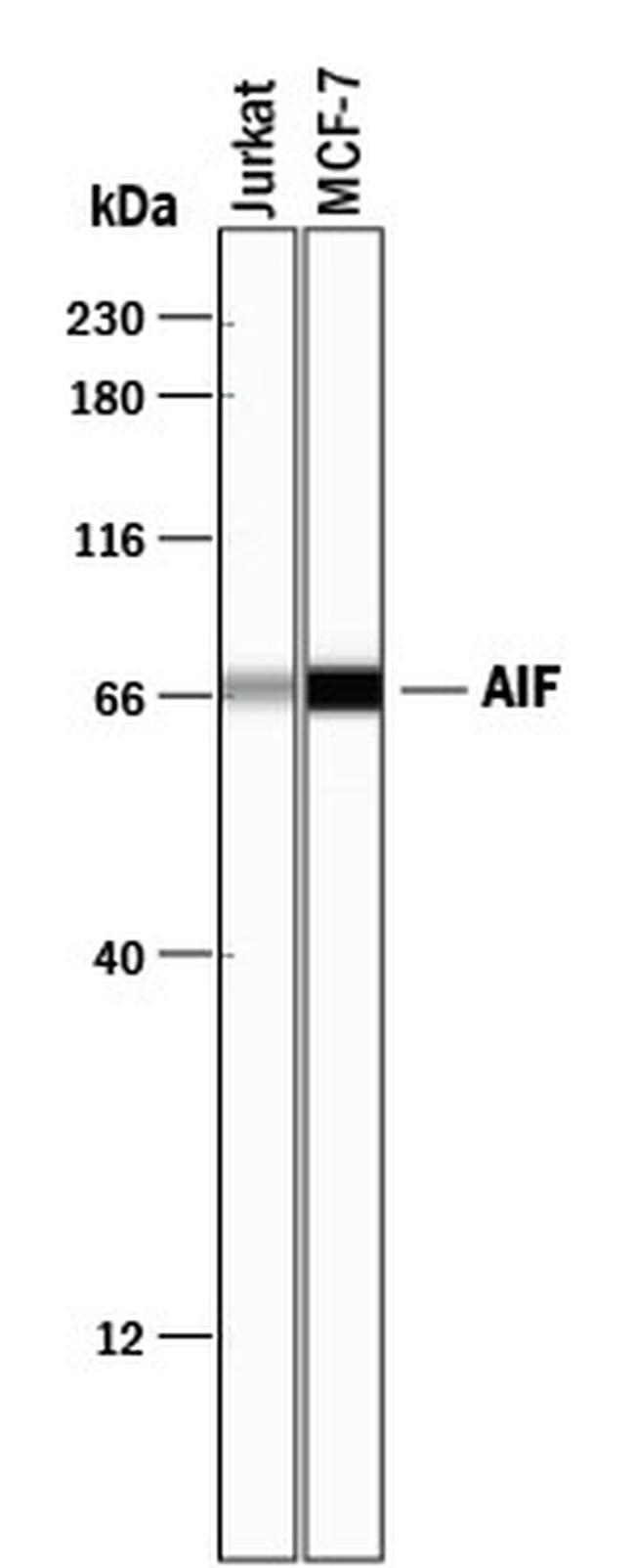
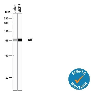
- Western blot analysis of AIF in 0.2 mg/mL lysates of Jurkat human acute T cell leukemia cell line and MCF‚7 human breast cancer cell line. Samples were incubated in AIF polyclonal antibody (Product # PA5-48092) using a dilution of 10 µg/mL followed by HRP-conjugated Anti-Sheep IgG at a dilution of 0.0763888888888889. A specific band was detected for AIF at approximately 69 kDa (as indicated). This experiment was conducted under reducing conditions and using the 12-230 kDa separation system.
Supportive validation
- Submitted by
- Invitrogen Antibodies (provider)
- Main image
- Experimental details
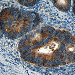
- Immunohistochemical analysis of AIF in immersion fixed paraffin-embedded sections of human colon. Samples were incubated in AIF polyclonal antibody (Product # PA5-48092) using a dilution of 10 µg/mL overnight at 4 °C. Before incubation with the primary antibody tissue was subjected to heat-induced epitope retrieval using Antigen Retrieval Reagent-Basic. Tissue was stained using the Anti-Sheep HRP-DAB Cell & Tissue Staining Kit (brown) and counterstained with hematoxylin (blue).
