Explore
 Validate
 Learn
Learn Western blot
 ELISA
ELISAAntibody data
- Antibody Data
- Antigen structure
- References [0]
- Comments [0]
- Validations
- Western blot [2]
- Immunohistochemistry [1]
Submit
Validation data
Reference
Comment
Report error
- Product number
- GTX22096 - Provider product page

- Provider
- GeneTex
- Proper citation
- GeneTex Cat#GTX22096, RRID:AB_369071
- Product name
- Cyclin B1 antibody
- Antibody type
- Polyclonal
- Reactivity
- Human, Mouse, Rat
- Host
- Rabbit
No comments: Submit comment
Supportive validation
- Submitted by
- GeneTex (provider)
- Main image
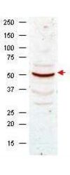
- Experimental details
- Western blot analysis using GeneTex Anti-Cyclin B1 antibody (GTX22096) shows detection of Cyclin B1 present in asynchronous HeLa cell lysates. Comparison to a molecular weight marker indicates a band of ~55 kDa corresponding to human Cyclin B1 (arrowhead). Approximately 50 ?g of lysate was loaded on to a 7% SDS-PAGE gel for separation. After transfer to nitrocellulose, the blot was incubated with a 1:500 dilution of the antibody for 1 h at room temperature. Detection occurred using a 1:10,000 of HRP conjugated Goat anti Rabbit IgG (GTX27090).
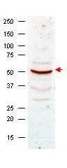
- Submitted by
- GeneTex (provider)
- Main image
- Experimental details
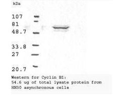
- Western blot analysis using GeneTex Anti-Cyclin B1 antibody (GTX22096) shows detection of human Cyclin B1 present in asynchronous HN30 cell lysates. HN30 cells are from head and neck cancer tumors that over express cyclin B1 and D1. Comparison to a molecular weight marker indicates a band of ~62 kDa corresponding to the expected molecular weight for the protein. The blot was incubated with a 1:500 dilution of the antibody for 1 h at room temperature. Detection occurred using a 1:10,000 of HRP conjugated Goat anti Rabbit IgG GTX27090 and chemiluminescence reagent with a 1min exposure time.

Supportive validation
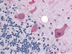
- Submitted by
- GeneTex (provider)
- Main image

- Experimental details
- GeneTex anti-Cyclin B1 antibody (GTX22096) was diluted 1:500 to detect Cyclin B1 in human brain cerebellum tissue. Tissue was formalin fixed and paraffin embedded. No pre-treatment of sample was required. The image shows the localization of antibody as the precipitated red signal, with a hematoxylin purple nuclear counter stain.