Explore
 Validate
 Learn
Learn710524
antibody from Invitrogen Antibodies
Targeting: EPCAM
17-1A, 323/A3, CD326, CO-17A, EGP-2, EGP34, EGP40, Ep-CAM, ESA, GA733-2, HEA125, KS1/4, KSA, Ly74, M4S1, MH99, MIC18, MK-1, MOC31, TACST-1, TACSTD1, TROP1
 Western blot
Antibody data
- Antibody Data
- Antigen structure
- References [1]
- Comments [0]
- Validations
- Western blot [4]
- Immunocytochemistry [1]
Submit
Validation data
Reference
Comment
Report error
- Product number
- 710524 - Provider product page

- Provider
- Invitrogen Antibodies
- Product name
- EpCAM Recombinant Polyclonal Antibody (22 HCLC)
- Antibody type
- Polyclonal
- Antigen
- Other
- Description
- This antibody is predicted to react with bovine, non-human primate, rabbit and ovine based on sequence homology.
- Antibody clone number
- 22 HCLC
- Concentration
- 0.5 mg/mL
Submitted references A reproducible scaffold-free 3D organoid model to study neoplastic progression in breast cancer.
Djomehri SI, Burman B, Gonzalez ME, Takayama S, Kleer CG
Journal of cell communication and signaling 2019 Mar;13(1):129-143
Journal of cell communication and signaling 2019 Mar;13(1):129-143
No comments: Submit comment
Supportive validation
- Submitted by
- Invitrogen Antibodies (provider)
- Main image

- Experimental details
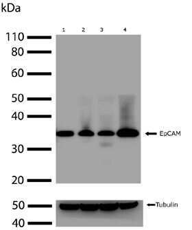
- Western blot analysis of endogenous EpCAM in 30 µg of HeLa (lane 1), MDAMB231 (lane 2), HEK-293 (lane 3), and COS-7 (lane 4) whole cell extracts using an EpCAM Recombinant Rabbit Polyclonal Antibody (Product # 710524) at a concentration of 2 µg/mL. A rabbit Anti-tubulin antibody was blotted as a loading control and the blots were developed using chemiluminescence (ECL) method with a Goat anti-Rabbit HRP secondary antibody (Product # G-21234). Results show a band at ~35kDa.
- Submitted by
- Invitrogen Antibodies (provider)
- Main image
- Experimental details
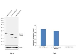
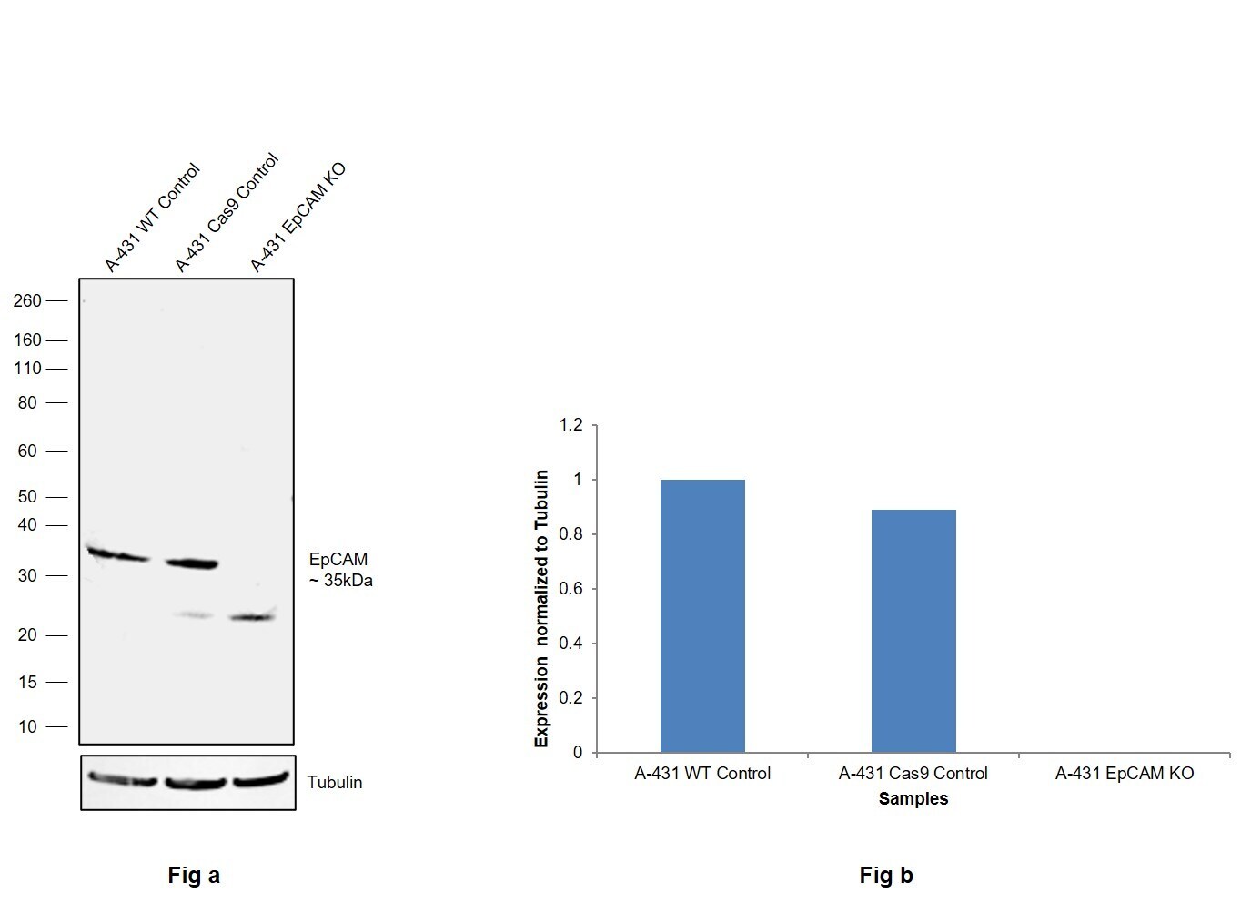
- Knockout of EpCAM was achieved by CRISPR-Cas9 genome editing using LentiArray™ Lentiviral sgRNA (Product # A32042, Assay ID CRISPR701274_LV) and LentiArray Cas9 Lentivirus (Product # A32064). Western blot analysis of EpCAM was performed by loading 30 µg of A-431 Wild Type (Lane 1), A-431 Cas9 (Lane 2) andA-431 EpCAM KO (Lane 3) membrane enriched extracts. The samples were electrophoresed using NuPAGE™ Novex™ 4-12% Bis-Tris Protein Gel (Product # NP0322BOX). Resolved proteins were then transferred onto a PVDF membrane (Product # LC2002) by iBlot® 2 Dry Blotting System (Product # IB21001). The blot was probed with an EpCAM Recombinant Polyclonal Antibody (22 HCLC) (Product # 710524, 1:1,000 dilution) and Goat anti-Rabbit IgG (H+L) Superclonal™ Recombinant Secondary Antibody, HRP (Product # A27036, 1:6,000 dilution) using the iBright FL 1000 (Product # A32752). Chemiluminescent detection was performed using SuperSignal™ West Dura Extended Duration Substrate (Product # 34076). Loss of signal upon CRISPR mediated knockout (KO) using the LentiArray™ CRISPR product line confirms that antibody is specific to EpCAM. An uncharacterized band was observed in CAS9 and EpCAM KO samles at ~25 kDa.
- Submitted by
- Invitrogen Antibodies (provider)
- Main image
- Experimental details
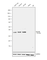
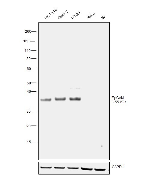
- Western Blot was performed using Anti-EpCAM Recombinant Polyclonal Antibody (22 HCLC) (Product # 710524) and a 35 kDa band corresponding to Epithelial cell adhesion molecule was observed across cell lines tested. Membrane enriched extracts (30 µg lysate) of HCT 116 (Lane 1), Caco-2 (Lane 2), HT-29 (Lane 3), HeLa (Lane 4) and BJ (Lane 5) were electrophoresed using NuPAGE™ 4-12% Bis-Tris Protein Gel (Product # NP0321BOX). Resolved proteins were then transferred onto a nitrocellulose membrane (Product # IB23001) by iBlot® 2 Dry Blotting System (Product # IB21001). The blot was probed with the primary antibody (1:1000 dilution) and detected by chemiluminescence with Goat anti-Rabbit IgG (H+L) Superclonal™ Recombinant Secondary Antibody, HRP (Product # A27036, 1:20000 dilution) using the iBright FL 1000 (Product # A32752). Chemiluminescent detection was performed using SuperSignal™ West Pico PLUS Chemiluminescent Substrate (Product # 34580).
- Submitted by
- Invitrogen Antibodies (provider)
- Main image
- Experimental details
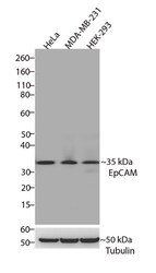
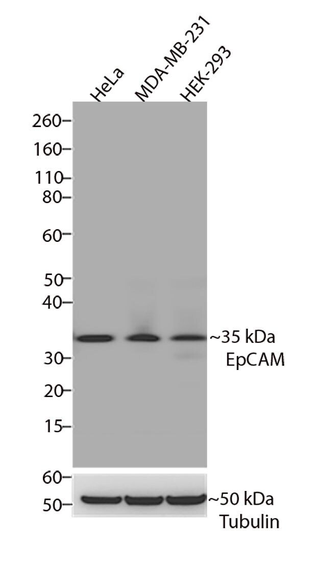
- Western blot analysis of EpCAM/CD326 was performed by loading 30 µg of HeLa, MDA-MB-231 and HEK-293 cell lysates using Novex®NuPAGE® 4-12% Bis-Tris gel (Product # NP0321BOX), XCell SureLock Electrophoresis System (Product # EI0002), Novex® Sharp Pre-Stained Protein Standard (Product # LC5800), and iBlot® Dry Blotting System (Product # IB21001). Proteins were transferred to a nitrocellulose membrane and blocked with 5% skim milk for 1 hour at room temperature. EpCAM/CD326 was detected at ~35 kDa using EpCAM/CD326 Recombinant Rabbit Polyclonal Antibody (Product # 710524) at a 1:1000 dilution in 2.5% skim milk at 4°C overnight on a rocking platform. Detection was performed using an HRP-conjugated Goat anti-Rabbit secondary antibody (Product # G-21234) at a 1:5000 dilution and chemiluminescent detection was performed using Pierce™ ECL Western blotting Substrate (Product # 32106).
Supportive validation
- Submitted by
- Invitrogen Antibodies (provider)
- Main image
- Experimental details
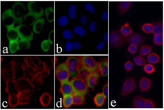
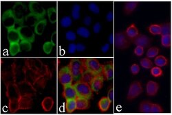
- Immunofluorescent analysis of EpCAM/CD326 was performed on 70% confluent log phase HeLa cells. The cells were fixed with 4% paraformaldehyde for 15 minutes, permeabilized with 0. 25% Triton X-100 for 10 minutes, and blocked with 5% BSA for 1 hour at room temperature. The cells were labeled with EpCAM/CD326 Recombinant Rabbit Polyclonal Antibody (Product # 710524) at a dilution of 1:500 in 1% BSA and incubated for 3 hours at room temperature and then labeled with Alexa Fluor® 488 Goat anti-Rabbit IgG secondary antibody (Product # A-11008) at a dilution of 1:400 for 30 minutes at room temperature (Panel a: green). Nuclei (Panel b: blue) were stained with SlowFade® Gold Antifade Mountant with DAPI (Product # S36938). F-actin (Panel c: red) was stained with Alexa Fluor 594 phalloidin (Product # A12381). Panel d is a merged image showing cytoplasmic localization and panel e is a control without primary antibody. The images were captured using a Nikon microscope at 20X magnification.