Explore
 Validate
 Learn
Learn53-8326-42
antibody from Invitrogen Antibodies
Targeting: EPCAM
17-1A, 323/A3, CD326, CO-17A, EGP-2, EGP34, EGP40, Ep-CAM, ESA, GA733-2, HEA125, KS1/4, KSA, Ly74, M4S1, MH99, MIC18, MK-1, MOC31, TACST-1, TACSTD1, TROP1
 Flow cytometry
Antibody data
- Antibody Data
- Antigen structure
- References [9]
- Comments [0]
- Validations
- Flow cytometry [1]
- Other assay [2]
Submit
Validation data
Reference
Comment
Report error
- Product number
- 53-8326-42 - Provider product page

- Provider
- Invitrogen Antibodies
- Product name
- CD326 (EpCAM) Monoclonal Antibody (MH99), Alexa Fluor™ 488, eBioscience™
- Antibody type
- Monoclonal
- Antigen
- Other
- Description
- Description: The monoclonal antibody MH99 recognizes human CD326 also known as EpCAM (Epithelial cell adhesion molecule). This 40 kDa type I transmembrane protein is involved in cell-cell interactions in a calcium-independent manner. EpCAM is expressed primarily on the basolateral surface of most epithelia. Although normal epithelia express low levels of CD326, increased expression has been correlated with increased proliferation and progression to a mesenchymal phenotype. CD326 has also been used as a diagnostic marker on circulating metastatic carcinoma cells, while cancer cells of non-epithelial origin do not express EpCAM.
- Conjugate
- Green dye
- Antibody clone number
- MH99
- Concentration
- 5 µL/Test
Submitted references A human multi-cellular model shows how platelets drive production of diseased extracellular matrix and tissue invasion.
Modelling TGFβR and Hh pathway regulation of prognostic matrisome molecules in ovarian cancer.
Mechanical Stimulation Modulates Osteocyte Regulation of Cancer Cell Phenotype.
Profiling protein expression in circulating tumour cells using microfluidic western blotting.
Expression of epithelial cell adhesion molecule in carcinoma cells present in blood and primary and metastatic tumors.
Selection of carbohydrate antigens in human epithelial ovarian cancers as targets for immunotherapy: serous and mucinous tumors exhibit distinctive patterns of expression.
Evidence for a role of the epithelial glycoprotein 40 (Ep-CAM) in epithelial cell-cell adhesion.
Ep-CAM: a human epithelial antigen is a homophilic cell-cell adhesion molecule.
Monoclonal antibodies to three widely distributed human cell surface antigens.
Malacrida B, Nichols S, Maniati E, Jones R, Delanie-Smith R, Roozitalab R, Tyler EJ, Thomas M, Boot G, Mackerodt J, Lockley M, Knight MM, Balkwill FR, Pearce OMT
iScience 2021 Jun 25;24(6):102676
iScience 2021 Jun 25;24(6):102676
Modelling TGFβR and Hh pathway regulation of prognostic matrisome molecules in ovarian cancer.
Delaine-Smith RM, Maniati E, Malacrida B, Nichols S, Roozitalab R, Jones RR, Lecker LSM, Pearce OMT, Knight MM, Balkwill FR
iScience 2021 Jun 25;24(6):102674
iScience 2021 Jun 25;24(6):102674
Mechanical Stimulation Modulates Osteocyte Regulation of Cancer Cell Phenotype.
Verbruggen SW, Thompson CL, Duffy MP, Lunetto S, Nolan J, Pearce OMT, Jacobs CR, Knight MM
Cancers 2021 Jun 10;13(12)
Cancers 2021 Jun 10;13(12)
Profiling protein expression in circulating tumour cells using microfluidic western blotting.
Sinkala E, Sollier-Christen E, Renier C, Rosàs-Canyelles E, Che J, Heirich K, Duncombe TA, Vlassakis J, Yamauchi KA, Huang H, Jeffrey SS, Herr AE
Nature communications 2017 Mar 23;8:14622
Nature communications 2017 Mar 23;8:14622
Expression of epithelial cell adhesion molecule in carcinoma cells present in blood and primary and metastatic tumors.
Rao CG, Chianese D, Doyle GV, Miller MC, Russell T, Sanders RA Jr, Terstappen LW
International journal of oncology 2005 Jul;27(1):49-57
International journal of oncology 2005 Jul;27(1):49-57
Selection of carbohydrate antigens in human epithelial ovarian cancers as targets for immunotherapy: serous and mucinous tumors exhibit distinctive patterns of expression.
Federici MF, Kudryashov V, Saigo PE, Finstad CL, Lloyd KO
International journal of cancer 1999 Apr 12;81(2):193-8
International journal of cancer 1999 Apr 12;81(2):193-8
Evidence for a role of the epithelial glycoprotein 40 (Ep-CAM) in epithelial cell-cell adhesion.
Litvinov SV, Bakker HA, Gourevitch MM, Velders MP, Warnaar SO
Cell adhesion and communication 1994 Oct;2(5):417-28
Cell adhesion and communication 1994 Oct;2(5):417-28
Ep-CAM: a human epithelial antigen is a homophilic cell-cell adhesion molecule.
Litvinov SV, Velders MP, Bakker HA, Fleuren GJ, Warnaar SO
The Journal of cell biology 1994 Apr;125(2):437-46
The Journal of cell biology 1994 Apr;125(2):437-46
Monoclonal antibodies to three widely distributed human cell surface antigens.
Mattes MJ, Cairncross JG, Old LJ, Lloyd KO
Hybridoma 1983;2(3):253-64
Hybridoma 1983;2(3):253-64
No comments: Submit comment
Supportive validation
- Submitted by
- Invitrogen Antibodies (provider)
- Main image
- Experimental details
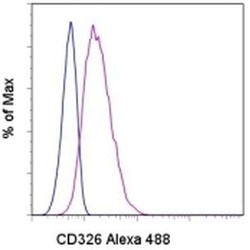
- Staining of A549 cells with Mouse IgG2a K Isotype Control Alexa Fluor® 488 (Product # 53-4724-80) (blue histogram) or Anti-Human CD326 (EpCAM) Alexa Fluor® 488 (purple histogram). Total viable cells were used for analysis.
- Conjugate
- Green dye

Supportive validation
- Submitted by
- Invitrogen Antibodies (provider)
- Main image
- Experimental details
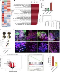
- Figure 7 TGFbetaR and GLI1/2 inhibitor combination reduces tumor matrix expression in HGSOC tri-cultures RNAseq was performed on adipocyte-only gels (Adipo_only), control tri-cultures (G164Tri), G164 + OF co-cultures, and G164Tri treated with inhibitor combination (G164Tri + CombI). (A) Heatmap of all matrisome genes detected in Adipo_only, G164 + OF, and G164Tri. (B) GSEA was performed on differentially expressed genes in G164Tri vs G164 + OF; bar plot indicates normalized enrichment scores (NES) for the indicated gene ontologies (p < 0.05). (C) Matrix index was calculated from the RNAseq data across the samples. (D) Hierarchical cluster analysis of transcriptomes for indicated samples. (E) After 21 days of culture, gel areas of control and combination-treated tri-cultures were measured from images (insets show 2 different experiments); data are mean of 2-4 gels per experiment (N = 5), p < 0.05 (two-way, paired t test). (F and G) (F) IF on tri-cultures for EPCAM, alphaSMA, FN1, VCAN, COL1A1, COMP, COL11A1, and DAPI (blue), scale bars represent 100 mum. G164Tri was treated with inhibitor combination and subjected to RNAseq (G) Volcano plot used to visualize changes in expression between G164Tri treated with inhibitors (G164Tri + CombI) and control G164Tri. Red dots indicate adjusted P value
- Conjugate
- Green dye

- Submitted by
- Invitrogen Antibodies (provider)
- Main image
- Experimental details
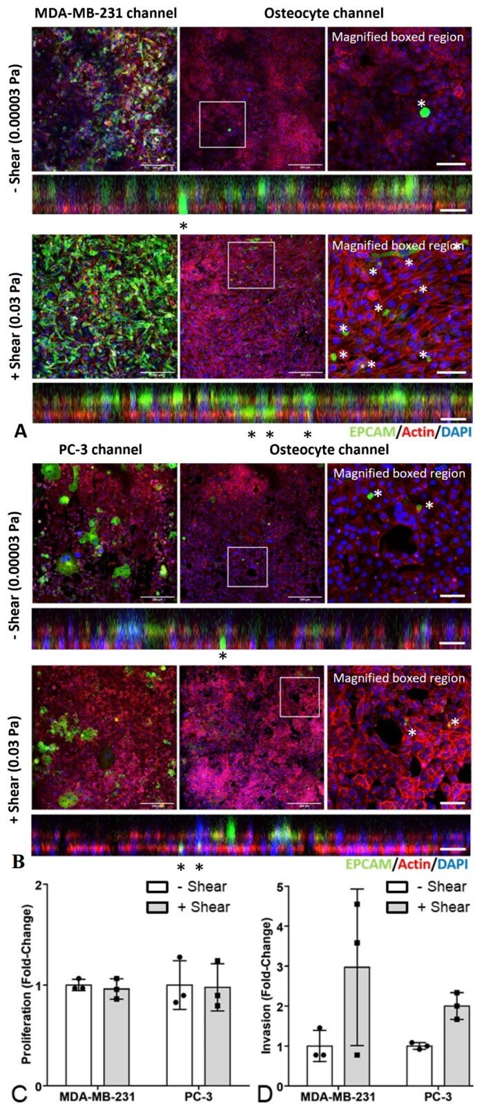
- Figure 5 Mechanical stimulation of osteocytes had no effect on proliferation but increased invasion in organ-chip models of breast and prostate cancer bone metastasis. Representative confocal images of ( A ) the MDA-MB-231 breast cancer bone metastasis chip and ( B ) the PC-3 prostate cancer bone metastasis chip. Immunofluorescent staining for cancer cells (EpCAM, green), actin (Phalloidin, red) and nuclei (DAPI, blue) in both the cancer cell channel and the osteocyte channel. The images are single confocal planes with an additional magnified view of the osteocyte channel (right) and the orthogonal projection showing both channels (below). The upper panels show images from organ chips in which the MLO-Y4 cells were exposed to minimal shear stress (0.00003 Pa), with lower panels showing organ-chips with higher shear conditions (0.03 Pa). Invasion of EpCAM-stained cancer cells through the porous membrane into the bone channel are indicated (*). Scale bar = 20 um. Associated analysis of EpCAM-positive cancer cells to quantify ( C ) proliferation in both channels, and ( D ) invasion into the bone channel. Data based on three separate confocal z-stacks for each chip.
- Conjugate
- Green dye