Explore
 Validate
 Learn
Learn Western blot
Antibody data
- Antibody Data
- Antigen structure
- References [1]
- Comments [0]
- Validations
- Western blot [1]
- Other assay [1]
Submit
Validation data
Reference
Comment
Report error
- Product number
- MA5-15162 - Provider product page

- Provider
- Invitrogen Antibodies
- Product name
- MEK1/MEK2 Monoclonal Antibody (E.901.7)
- Antibody type
- Monoclonal
- Antigen
- Synthetic peptide
- Description
- It is not recommended to aliquot this antibody.
- Reactivity
- Human, Mouse, Rat, Canine
- Host
- Rabbit
- Isotype
- IgG
- Antibody clone number
- E.901.7
- Vial size
- 100 µL
- Storage
- -20°C
Submitted references CRMP2 Is Involved in Regulation of Mitochondrial Morphology and Motility in Neurons.
Brustovetsky T, Khanna R, Brustovetsky N
Cells 2021 Oct 17;10(10)
Cells 2021 Oct 17;10(10)
No comments: Submit comment
Supportive validation
- Submitted by
- Invitrogen Antibodies (provider)
- Main image
- Experimental details
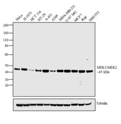
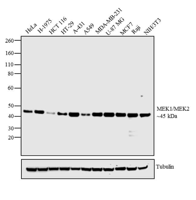
- Western blot analysis was performed on whole cell extracts (30 µg lysate) of HeLa (Lane 1), H-1975 (Lane 2), HCT 116 (Lane 3), HT-29 (Lane 4), A-431 (Lane 5), A549 (Lane 6) MDA-MB-231 (Lane 7), U-87 MG (Lane 8), MCF7 (Lane 9), Raji (Lane 10) and NIH/3T3 (Lane 11). The blot was probed with Anti-MEK1/MEK2 Rabbit Monoclonal Antibody (Product # MA5-15162, 1:1000 dilution) and detected by chemiluminescence using Goat anti-Rabbit IgG (H+L) Superclonal™ Secondary Antibody, HRP conjugate (Product # A27036, 0.25 µg/mL, 1:4000 dilution). A 45 kDa band corresponding to MEK1/MEK2 was detected across all cell lines tested. Known quantity of protein samples were electrophoresed using Novex® NuPAGE® 4-12 % Bis-Tris gel (Product # NP0321BOX), XCell SureLock™ Electrophoresis System (Product # EI0002) and Novex® Sharp Pre-Stained Protein Standard (Product # LC5800). Resolved proteins were then transferred onto a nitrocellulose membrane with iBlot® 2 Dry Blotting System (Product # IB21001). The membrane was probed with the relevant primary and secondary Antibody following blocking with 5 % skimmed milk. Chemiluminescent detection was performed using Pierce™ ECL Western Blotting Substrate (Product # 32106).
Supportive validation
- Submitted by
- Invitrogen Antibodies (provider)
- Main image

- Experimental details
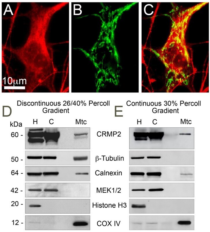
- Figure 1 Interaction of CRMP2 with brain mitochondria. ( A - C ) Immunocytochemistry staining with anti-CRMP2 antibody ( A , red) and visualization of mitochondria with mitochondrially targeted enhanced yellow fluorescent protein (mito-eYFP) ( B , green) illustrates co-localization of CRMP2 and mitochondria (yellow, C ) in cultured striatal neuron from FVB/NJ mouse. ( D , E ), CRMP2 binds to synaptic brain mitochondria isolated from FVB/NJ mice. Mitochondria were purified either on discontinuous 26/40% Percoll gradient ( D ) or on continuous 30% Percoll gradient ( E ) as described in the Materials and Methods. H: homogenate; C: cytosolic fraction; Mtc: mitochondrial fraction. MEK1/2, calnexin, beta-tubulin, Histone H3, and COX IV are cytosolic, ER, microtubule, nuclear, and mitochondrial markers, respectively. Representative western blots from 3 independent experiments are shown.