Explore
 Validate
 Learn
Learn701064
antibody from Invitrogen Antibodies
Targeting: RPS6KB1
p70(S6K)-alpha, PS6K, S6K, S6K1, STK14A
 Western blot
Antibody data
- Antibody Data
- Antigen structure
- References [0]
- Comments [0]
- Validations
- Western blot [2]
- Immunocytochemistry [1]
- Flow cytometry [1]
Submit
Validation data
Reference
Comment
Report error
- Product number
- 701064 - Provider product page

- Provider
- Invitrogen Antibodies
- Product name
- Phospho-p70 S6 Kinase (Thr389) Recombinant Rabbit Monoclonal Antibody (B2H9L2)
- Antibody type
- Monoclonal
- Antigen
- Synthetic peptide
- Description
- Intact IgG appears on a non-reducing gel as ~150 kDa band and upon reduction generating a ~25 kDa light chain band and a ~50 kDa heavy chain.
- Antibody clone number
- B2H9L2
- Concentration
- 0.5 mg/mL
No comments: Submit comment
Supportive validation
- Submitted by
- Invitrogen Antibodies (provider)
- Main image
- Experimental details
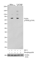
- Western blot analysis of p70S6K (pT389) was performed by loading 30 µg of untreated HeLa cells (lane 1) and HeLa cells treated with IGF-1 (150 ng/mL for 15 minutes) (lane 2) and U-87 MG lysates (lane 4) using Novex®NuPAGE®4-12% Bis-Tris gel (Product # NP0321BOX), XCell SureLock Electrophoresis System (Product # EI0002), and Novex® Sharp Pre-Stained Protein Standard (Product # LC5800). Proteins were transferred to a PVDF membrane and blocked with 5% skim milk for 1 hour at room temperature. p70S6K (pT389) was detected at ~70 kDa using p70S6K (pT389) Recombinant Rabbit Monoclonal Antibody (Product # 701064) at a 1:1000 dilution in 2.5% skim milk at 4°C overnight on a rocking platform. To confirm specificity, competition was performed with the phosphopeptide (10 µg/mL) (lane 3, 5). Detection was performed using an HRP-conjugated Goat anti-Rabbit secondary antibody (Product # G-21234) at a 1:5000 dilution and chemiluminescent detection was performed using Pierce™ ECL Western blotting Substrate (Product # 32106).

- Submitted by
- Invitrogen Antibodies (provider)
- Main image

- Experimental details
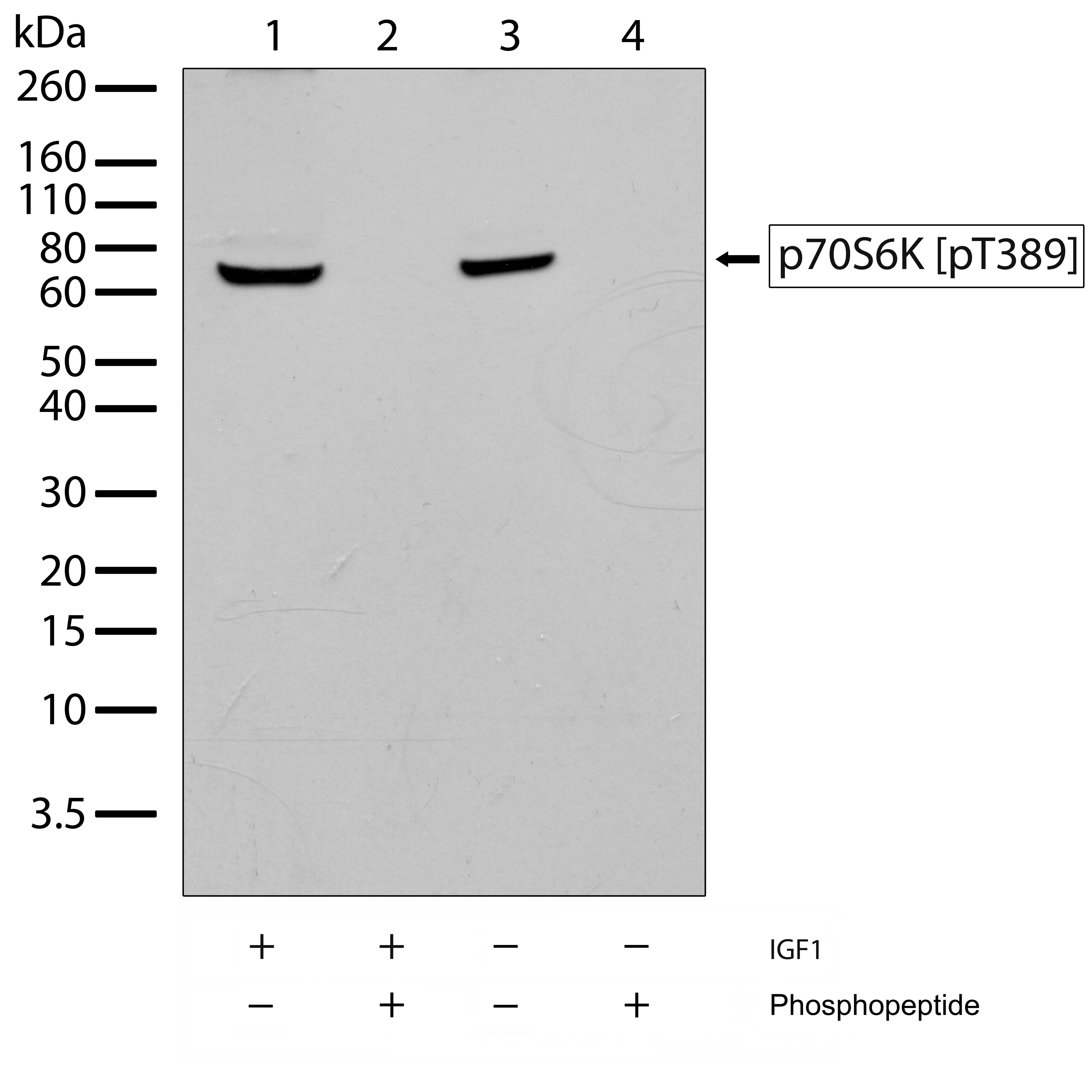
- Western blot analysis of Phospho-p70S6K Thr389 in whole cell extracts of serum-starved HeLa cells treated with IGF1 (150 ng/mL, 15 min) (lanes 1 and 2), and U87-MG cells (lanes 3 and 4) using a Phospho-p70S6K Thr389 recombinant rabbit monoclonal antibody (Product # 701064) at a dilution of 1 µg/mL. To confirm specificity, competition was performed by preincubation with the phosphopeptide to inhibit antibody binding (lanes 2 and 4). Samples were detected using chemiluminescence (ECL). Results show a band at ~70 kDa.
Supportive validation
- Submitted by
- Invitrogen Antibodies (provider)
- Main image
- Experimental details
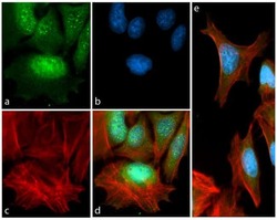
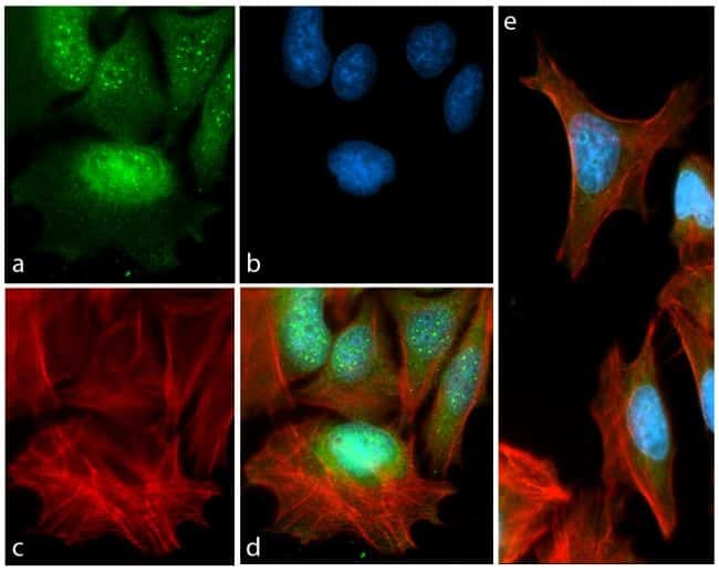
- Immunofluorescent analysis of Phospho-p70S6 Thr389 in serum-starved HeLa cells treated with insulin (100 ng/mL, 15 min) using a Phospho-p70S6 Thr389 recombinant rabbit monoclonal antibody (Product # 701064) followed by detection using an Alexa Fluor 488-conjugated goat anti-rabbit secondary antibody (green) (Image A). Nuclei were stained using DAPI (Image B) and actin stained with Alexa Fluor 594 phalloidin (red) (image C). Image D is a composite image showing cytoplasmic and nuclear localization of phosphorylated p70S6K and Image E is a composite image of cells showing competition with the phospho p70S6K (pT389) peptide.
Supportive validation
- Submitted by
- Invitrogen Antibodies (provider)
- Main image
- Experimental details
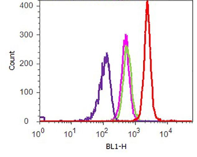
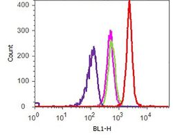
- Flow cytometry analysis of p70S6K (pT389) was performed on U-87 MG cells. Cells were fixed with 70% ethanol for 10 minutes, permeabilized with 0. 25% Tritonª X-100 for 20 minutes, and blocked with 5% BSA for 1 hour at room temperature. Cells were labeled with ABfinityª p70S6K (pT389) recombinant rabbit monoclonal antibody (Product # 701064, red histogram) or with rabbit isotype control (pink histogram) at a dilution of 1:250 in 2.5% BSA. After incubation at room temperature for 3 hours, the cells were labeled with Alexa Fluor¨ 488 goat anti-Rabbit Secondary antibody (Product # A11008) at a dilution of 1:400 for 30 minutes at room temperature. The representative 10,000 cells were acquired and analyzed for each sample using an Attune¨ Acoustic Focusing Cytometer. The purple histogram represents unstained control cells and the green histogram represents no-primary-antibody control.