 Explore
 Validate
 Learn
Learn Western blot
Antibody data
- Antibody Data
- Antigen structure
- References [1]
- Comments [0]
- Validations
- Western blot [3]
- Immunocytochemistry [1]
- Immunohistochemistry [2]
Submit
Validation data
Reference
Comment
Report error
- Product number
- PA5-16561 - Provider product page

- Provider
- Invitrogen Antibodies
- Product name
- PARP1 Polyclonal Antibody
- Antibody type
- Polyclonal
- Antigen
- Synthetic peptide
- Description
- PA5-16561 was successfully used to detect PARP in WB, IHC and IF applications.
- Concentration
- 1 mg/mL
Submitted references Autophagy induced by SAHA affects mutant P53 degradation and cancer cell survival.
Foggetti G, Ottaggio L, Russo D, Mazzitelli C, Monti P, Degan P, Miele M, Fronza G, Menichini P
Bioscience reports 2019 Feb 28;39(2)
Bioscience reports 2019 Feb 28;39(2)
No comments: Submit comment
Supportive validation
- Submitted by
- Invitrogen Antibodies (provider)
- Main image
- Experimental details
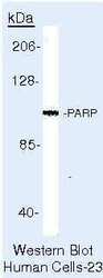
- Western blot of PARP (Poly ADP-Ribose Polymerase) using PARP (Poly ADP-Ribose Polymerase) Polyclonal Antibody (Product # PA5-16561) on Raji Cells.

- Submitted by
- Invitrogen Antibodies (provider)
- Main image

- Experimental details
- Western blot analysis of Poly ADP ribose Polymerase (PARP) was performed by loading 40 µg of Nuclear protein extracts from BEAS-2B (lane 1), HeLa (lane 2) Drosophila S2 cells (lane 3) in sample buffer and 5µL Spectra Multicolor Broad Range Protein Ladder (Product # 26634) onto a 10% polyacrylamide gel. Proteins were transferred to PVDF membrane (Product # 88518) at 65V for 90 min on ice. Membrane was blocked in 5% Milk in PBST for 1 hour at room temperature. PARP was detected at approximately 125 kDa using PARP polyclonal antibody (Product # PA5-16561) at a dilution of 1:1500 in 0.5% Milk in PBST overnight at 4°C. Followed by goat anti-rabbit alkaline phosphatase secondary antibody at a dilution of 1:20,000. Chemiluminescent detection was performed using ECF substrate. Data courtesy of Dr. Fondufe-Mittendorf's lab.

- Submitted by
- Invitrogen Antibodies (provider)
- Main image

- Experimental details
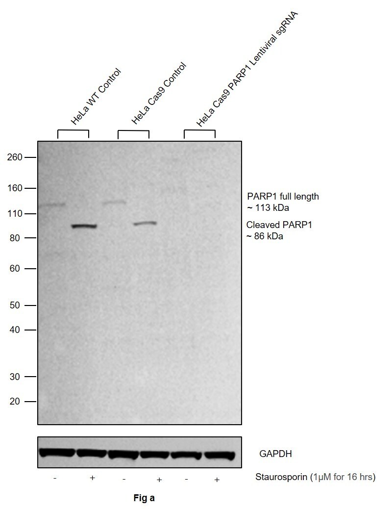
- CRISPR-Cas9 mediated genome editing ofPARP1 (as confirmed by next generation sequencing) was achieved by using LentiArray™ Lentiviral sgRNA (Product # A32042, AssayID CRISPR978664_LV) and LentiArray Cas9 Lentivirus (Product # A32064). Fig (a) Western blot analysis of PARP1 was performed by loading 30 µg of HeLa Wild Type (Lane 1), Treated HeLa Wild Type (Lane 2), HeLa Cas9 (Lane 3), Treated HeLa Cas9 (Lane 4), HeLa Cas9 cells transduced with PARP1 Lentiviral sgRNA (Lane 5) and Treated HeLa Cas9 cells transduced with PARP1 Lentiviral sgRNA (Lane 6) whole cell extracts. The samples were electrophoresed using NuPAGE™ Novex™ 4-12% Bis-Tris Protein Gel (Product # NP0322BOX). Resolved proteins were then transferred onto a nitrocellulose membrane (Product # IB23001) by iBlot® 2 Dry Blotting System (Product # IB21001). The blot was probed with Anti-PARP1 Polyclonal Antibody (Product # PA5-16561) using 1:1,500 dilution and Goat anti-Rabbit IgG (H+L) Superclonal™ Recombinant Secondary Antibody, HRP (Product # A27036 1:20,000 dilution).Chemiluminescent detection was performed using Novex® ECL Chemiluminescent Substrate Reagent Kit (Product # WP20005). Loss of signal in sgRNA transduced cells using the LentiArray™ CRISPR product line confirms that antibody is specific to PARP1. Treatment used is 1 µM Staurosporin for 16 hrs.
Supportive validation
- Submitted by
- Invitrogen Antibodies (provider)
- Main image
- Experimental details
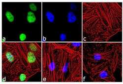
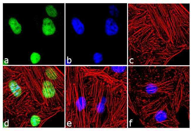
- Immunofluorescence analysis of PARP was performed using 70% confluent log phase HeLa cells treated with 1 uM staurosporine for 16 hours. The cells were fixed with 4% paraformaldehyde for 10 minutes, permeabilized with 0.1% Triton™ X-100 for 10 minutes, and blocked with 2% BSA for 1 hour at room temperature. The cells were labeled with PARP (Poly ADP-Ribose Polymerase) Rabbit Polyclonal Antibody (Product # PA5-16561) at 2 µg/mL in 0.1% BSA and incubated for 3 hours at room temperature and then labeled with Goat anti-Rabbit IgG (H+L) Superclonal™ Secondary Antibody, Alexa Fluor® 488 conjugate (Product # A27034) a dilution of 1:2000 for 45 minutes at room temperature (Panel a: green). Nuclei (Panel b: blue) were stained with SlowFade® Gold Antifade Mountant with DAPI (Product # S36938). F-actin (Panel c: red) was stained with Alexa Fluor® 555 Rhodamine Phalloidin (Product # R415, 1:300). Panel d represents the merged image showing nuclear localization. Panel e shows untreated cells with no signal. Panel f represents control cells with no primary antibody to assess background. The images were captured at 60X magnification.
Supportive validation
- Submitted by
- Invitrogen Antibodies (provider)
- Main image
- Experimental details
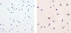
- Immunohistochemistry analysis of PARP (Poly ADP-Ribose Polymerase) showing staining in the nucleus of paraffin-embedded human brain tissue (right) compared to a negative control without primary antibody (left). To expose target proteins, antigen retrieval was performed using 10mM sodium citrate (pH 6.0), microwaved for 8-15 min. Following antigen retrieval, tissues were blocked in 3% H2O2-methanol for 15 min at room temperature, washed with ddH2O and PBS, and then probed with a PARP (Poly ADP-Ribose Polymerase) Rabbit Polyclonal Antibody (Product # PA5-16561) diluted in 3% BSA-PBS at a dilution of 1:50 for 1 hour at 37°C in a humidified chamber. Tissues were washed extensively in PBST and detection was performed using an HRP-conjugated secondary antibody followed by colorimetric detection using a DAB kit. Tissues were counterstained with hematoxylin and dehydrated with ethanol and xylene to prep for mounting.

- Submitted by
- Invitrogen Antibodies (provider)
- Main image
- Experimental details
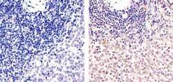
- Immunohistochemistry analysis of PARP (Poly ADP-Ribose Polymerase) showing staining in the nucleus of paraffin-embedded mouse spleen tissue (right) compared to a negative control without primary antibody (left). To expose target proteins, antigen retrieval was performed using 10mM sodium citrate (pH 6.0), microwaved for 8-15 min. Following antigen retrieval, tissues were blocked in 3% H2O2-methanol for 15 min at room temperature, washed with ddH2O and PBS, and then probed with a PARP (Poly ADP-Ribose Polymerase) Rabbit Polyclonal Antibody (Product # PA5-16561) diluted in 3% BSA-PBS at a dilution of 1:20 for 1 hour at 37°C in a humidified chamber. Tissues were washed extensively in PBST and detection was performed using an HRP-conjugated secondary antibody followed by colorimetric detection using a DAB kit. Tissues were counterstained with hematoxylin and dehydrated with ethanol and xylene to prep for mounting.
