Explore
 Validate
 Learn
Learn Western blot
 Immunoprecipitation
Antibody data
- Antibody Data
- Antigen structure
- References [1]
- Comments [0]
- Validations
- Western blot [4]
- Immunocytochemistry [1]
Submit
Validation data
Reference
Comment
Report error
- Product number
- MA5-18060 - Provider product page

- Provider
- Invitrogen Antibodies
- Product name
- PP2A alpha Monoclonal Antibody (7A6)
- Antibody type
- Monoclonal
- Antigen
- Synthetic peptide
- Description
- This antibody recognizes the C-terminal of PP2A, both alpha and beta isoforms, in human, mouse, rat and S.cerevisiae samples.
- Reactivity
- Human, Mouse, Rat, Yeast
- Host
- Mouse
- Isotype
- IgG
- Antibody clone number
- 7A6
- Vial size
- 100 µL
- Concentration
- 1 mg/mL
- Storage
- Maintain refrigerated at 2-8°C for up to 1 month. For long term storage store at -20°C
Submitted references Microhomology-based CRISPR tagging tools for protein tracking, purification, and depletion.
Lin DW, Chung BP, Huang JW, Wang X, Huang L, Kaiser P
The Journal of biological chemistry 2019 Jul 12;294(28):10877-10885
The Journal of biological chemistry 2019 Jul 12;294(28):10877-10885
No comments: Submit comment
Supportive validation
- Submitted by
- Invitrogen Antibodies (provider)
- Main image

- Experimental details
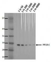
- Western blot analysis of PP2A in NIH-3T3 Puro lysates using a PPP2CA/PP2A alpha monoclonal antibody (Product # MA5-18060) at various dilutions, followed by detection using an AP-conjugated mouse IgG whole molecule antibody at a dilution of 1:2000 on a BCIP/NBT substrate.
- Submitted by
- Invitrogen Antibodies (provider)
- Main image
- Experimental details
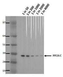
- Western blot analysis of PP2A in NIH-3T3 Puro lysates using a PPP2CA/PP2A alpha monoclonal antibody (Product # MA5-18060) at various dilutions, followed by detection using an AP-conjugated mouse IgG whole molecule antibody at a dilution of 1:2000 on a BCIP/NBT substrate.

- Submitted by
- Invitrogen Antibodies (provider)
- Main image
- Experimental details
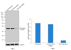
- Knockdown of PP2A alpha was achieved by transfecting PC-3 cells with PP2A alpha specific siRNAs (Silencer® select Product # s10958). Western blot analysis (Fig. a) was performed using whole cell extracts from the PP2A alpha knockdown cells (Lane 3), non-specific scrambled siRNA transfected cells (Lane 2) and untransfected cells (Lane 1). The blot was probed with Anti-PP2A alpha Monoclonal Antibody (7A6) (Product # MA5-18060, 1:1000 dilution) and Goat anti-Mouse IgG (H+L), Superclonal™ Recombinant Secondary Antibody, HRP (Product # A28177, 1:4000 dilution). Densitometric analysis of this Western Blot is shown in histogram (Fig. b). Decrease in signal upon siRNA mediated knock down confirms that antibody is specific to PP2A alpha.

- Submitted by
- Invitrogen Antibodies (provider)
- Main image

- Experimental details
- Western blot was performed using Anti-PP2A alpha Monoclonal Antibody (7A6) (Product # MA5-18060) and 36 kDa band corresponding to PP2A alpha was observed across the cell lines and tissue tested. Whole cell extracts (30 µg lysate) of A-431 (Lane1), PC-3 (Lane 2), HEL 92.1.7 (Lane 3), K-562 (Lane 4), A549 (Lane 5), HeLa (Lane 6) and tissue extracts (30 µg lysate) of Mouse Brain (Lane 7) were electrophoresed using Novex® NuPAGE® 4-12 % Bis-Tris gel (Product # NP0322BOX). Resolved proteins were then transferred onto a nitrocellulose membrane (Product # IB23001) by iBlot® 2 Dry Blotting System (Product # IB21001). The blot was probed with the primary antibody (1:1000 dilution) and detected by chemiluminescence with Goat anti-Mouse IgG (H+L), Superclonal™ Recombinant Secondary Antibody, HRP (Product # A28177, 1:4000 dilution) using the iBright FL 1000 (Product # A32752). Chemiluminescent detection was performed using Novex® ECL Chemiluminescent Substrate Reagent Kit (Product # WP20005).

Supportive validation
- Submitted by
- Invitrogen Antibodies (provider)
- Main image
- Experimental details
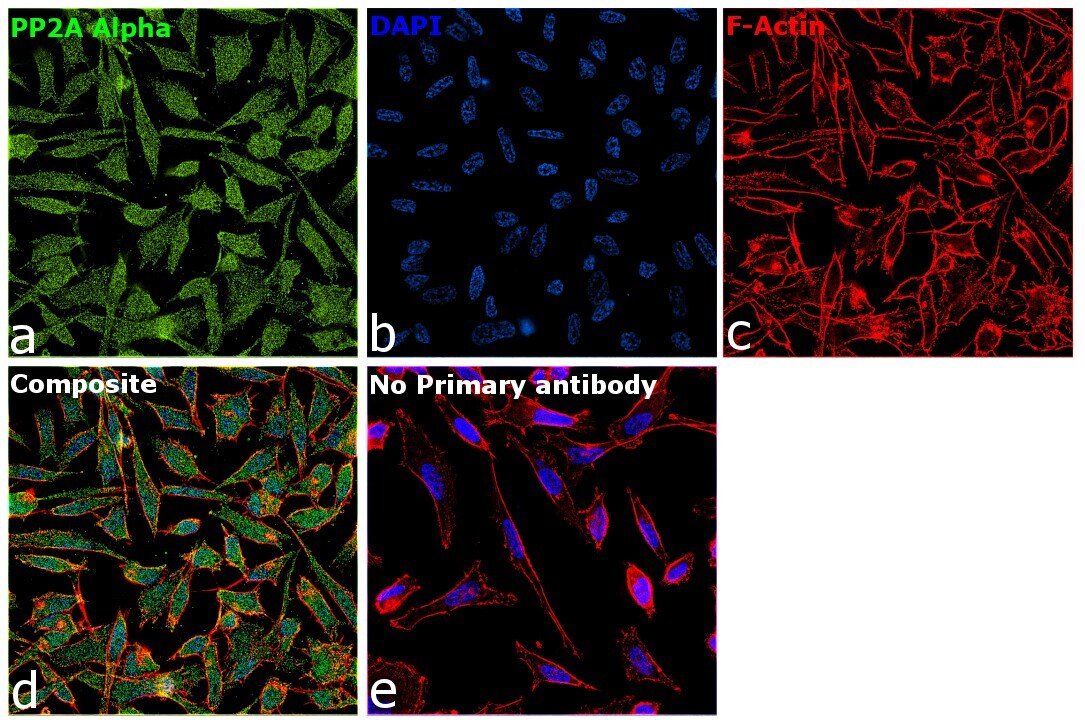
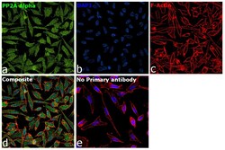
- Immunofluorescence analysis of PP2A alpha was performed using 70% confluent log phase PC-3 cells. The cells were fixed with 4% paraformaldehyde for 10 minutes, permeabilized with 0.1% Triton™ X-100 for 15 minutes, and blocked with 2% BSA for 1 hour at room temperature. The cells were labeled with PP2A alpha Monoclonal Antibody (7A6) (Product # MA5-18060) at 1:100 dilution in 0.1% BSA, incubated at 4 degree celsius overnight and then with Goat anti-Mouse IgG (H+L), Superclonal™ Recombinant Secondary Antibody, Alexa Fluor 488 (Product # A28175) at a dilution of 1:2000 for 45 minutes at room temperature (Panel a: Green). Nuclei (Panel b: Blue) were stained with SlowFade® Gold Antifade Mountant with DAPI (Product # S36938). F-actin (Panel c: Red) was stained with Rhodamine Phalloidin (Product # R415, 1:300). Panel d represents the merged image showing nuclear, cytoskeletal and cytoplasmic localization. Panel e represents control cells with no primary antibody to assess background. The images were captured at 60X magnification.