Explore
 Validate
 Learn
Learn Western blot
Antibody data
- Antibody Data
- Antigen structure
- References [1]
- Comments [0]
- Validations
- Western blot [1]
- Immunohistochemistry [2]
Submit
Validation data
Reference
Comment
Report error
- Product number
- 710087 - Provider product page

- Provider
- Invitrogen Antibodies
- Product name
- Phospho-PYK2 (Tyr402) Recombinant Polyclonal Antibody (17HCLC)
- Antibody type
- Polyclonal
- Antigen
- Other
- Description
- This antibody is predicted to react with mouse based on sequence homology.
- Antibody clone number
- 17HCLC
- Concentration
- 0.5 mg/mL
Submitted references The Tyrosine Kinase Pyk2 Contributes to Complement-Mediated Phagocytosis in Murine Macrophages.
Paone C, Rodrigues N, Ittner E, Santos C, Buntru A, Hauck CR
Journal of innate immunity 2016;8(5):437-51
Journal of innate immunity 2016;8(5):437-51
No comments: Submit comment
Supportive validation
- Submitted by
- Invitrogen Antibodies (provider)
- Main image

- Experimental details
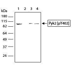
- Western blot analysis of Phospho-FAK2 pTyr402 in whole cell extract from A431 (50 µg/lane) (lane 1), samples pre-incubated with the phosphopeptide (0.5 µg) (lane 2), Jurkat cell lysate treated with PMA (5 ng/mL, 5 min) (lane 3), and Raji cell lysate (lane 4) using a Phospho-FAK2 pTyr402 Recombinant Rabbit Polyclonal Antibody (Product # 710087) at a dilution of 5 µg/mL.
Supportive validation
- Submitted by
- Invitrogen Antibodies (provider)
- Main image
- Experimental details
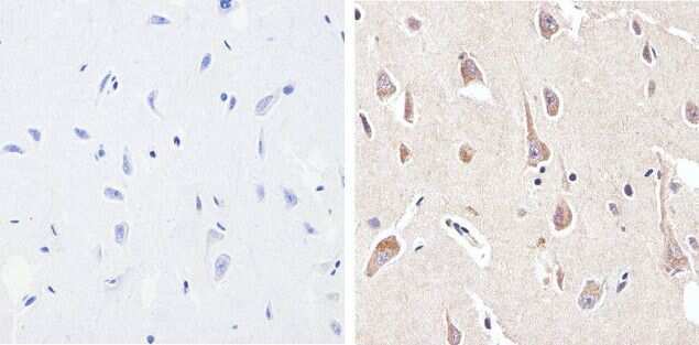
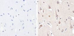
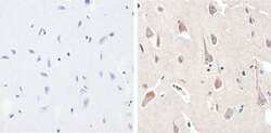
- Immunohistochemistry analysis of Phospho-FAK2 /PYK2 pTyr402 showing staining in the cytoplasm of paraffin-embedded human brain tissue (right) compared to a negative control without primary antibody (left). To expose target proteins, antigen retrieval was performed using 10 mM sodium citrate (pH 6.0), microwaved for 8-15 min. Following antigen retrieval, tissues were blocked in 3% H2O2-methanol for 15 min at room temperature, washed with ddH2O and PBS, and then probed with a Phospho-FAK2 /PYK2 pTyr402 (17HCLC) Recombinant Rabbit Polyclonal Antibody (Product # 710087) diluted in 3% BSA-PBS at a dilution of 1:20 overnight at 4°C in a humidified chamber. Tissues were washed extensively in PBST and detection was performed using a HRP-conjugated secondary antibody followed by colorimetric detection using a DAB kit. Tissues were counterstained with hematoxylin and dehydrated with ethanol and xylene to prep for mounting.

- Submitted by
- Invitrogen Antibodies (provider)
- Main image
- Experimental details
- Immunohistochemistry analysis of Phospho-FAK2 /PYK2 pTyr402 showing staining in the cytoplasm of paraffin-embedded human brain tissue (right) compared to a negative control without primary antibody (left). To expose target proteins, antigen retrieval was performed using 10 mM sodium citrate (pH 6.0), microwaved for 8-15 min. Following antigen retrieval, tissues were blocked in 3% H2O2-methanol for 15 min at room temperature, washed with ddH2O and PBS, and then probed with a Phospho-FAK2 /PYK2 pTyr402 (17HCLC) Recombinant Rabbit Polyclonal Antibody (Product # 710087) diluted in 3% BSA-PBS at a dilution of 1:20 overnight at 4°C in a humidified chamber. Tissues were washed extensively in PBST and detection was performed using a HRP-conjugated secondary antibody followed by colorimetric detection using a DAB kit. Tissues were counterstained with hematoxylin and dehydrated with ethanol and xylene to prep for mounting.