 Explore
 Validate
 Learn
Learn Western blot
Antibody data
- Antibody Data
- Antigen structure
- References [6]
- Comments [0]
- Validations
- Western blot [1]
- Immunohistochemistry [1]
- Other assay [1]
Submit
Validation data
Reference
Comment
Report error
- Product number
- 14-9864-82 - Provider product page

- Provider
- Invitrogen Antibodies
- Product name
- Phospho-NFkB p65 (Ser529) Monoclonal Antibody (MCFA30), eBioscience™
- Antibody type
- Monoclonal
- Antigen
- Other
- Description
- Description: The MCFA30 monoclonal antibody recognizes human NF kappa B (NFkB) p65 subunit when serine 529 is phosphorylated. NFkB, also known as nuclear factor kappa-light chain enhancer of activated B cells, is a ubiquitous transcription factor that regulates the transcription of many genes involved in cell proliferation, apoptosis, development, immunity and cancer. Functional NFkB is a homo- or hetero-dimer composed of 5 members of the NFkB family: p65 (RelA), c-Rel, RelB, p50 (NFkB1, p105 precursor protein), and p52 (NFkB2, p100 precursor protein). The activity of the complex is negatively regulated by binding to IkB inhibitors that sequester NFkB into the cytoplasm, inhibiting its transcriptional activity. NFkB-activating agents like tumor necrosis factor (TNF) alpha, interleukin-1 beta, lipopolysaccharide, camptothecin, and phorbol ester (PMA) induce the phosphorylation and degradation of IkB, leading to the translocation of NFkB to the nucleus where it binds to kB motifs and regulates gene expression. The activity of p65-containing NFkB complexes is positively regulated by phosphorylation of the p65 subunit at serine 529.
- Antibody clone number
- MCFA30
- Concentration
- 0.5 mg/mL
Submitted references Neutrophils induce macrophage anti-inflammatory reprogramming by suppressing NF-κB activation.
GITR intrinsically sustains early type 1 and late follicular helper CD4 T cell accumulation to control a chronic viral infection.
Activation of nuclear factor kappa B by different agents: influence of culture conditions in a cell-based assay.
Control of lymphocyte development by nuclear factor-kappaB.
Tumor necrosis factor alpha-induced phosphorylation of RelA/p65 on Ser529 is controlled by casein kinase II.
Function and activation of NF-kappa B in the immune system.
Marwick JA, Mills R, Kay O, Michail K, Stephen J, Rossi AG, Dransfield I, Hirani N
Cell death & disease 2018 Jun 4;9(6):665
Cell death & disease 2018 Jun 4;9(6):665
GITR intrinsically sustains early type 1 and late follicular helper CD4 T cell accumulation to control a chronic viral infection.
Clouthier DL, Zhou AC, Wortzman ME, Luft O, Levy GA, Watts TH
PLoS pathogens 2015 Jan;11(1):e1004517
PLoS pathogens 2015 Jan;11(1):e1004517
Activation of nuclear factor kappa B by different agents: influence of culture conditions in a cell-based assay.
Hellweg CE, Arenz A, Bogner S, Schmitz C, Baumstark-Khan C
Annals of the New York Academy of Sciences 2006 Dec;1091:191-204
Annals of the New York Academy of Sciences 2006 Dec;1091:191-204
Control of lymphocyte development by nuclear factor-kappaB.
Siebenlist U, Brown K, Claudio E
Nature reviews. Immunology 2005 Jun;5(6):435-45
Nature reviews. Immunology 2005 Jun;5(6):435-45
Tumor necrosis factor alpha-induced phosphorylation of RelA/p65 on Ser529 is controlled by casein kinase II.
Wang D, Westerheide SD, Hanson JL, Baldwin AS Jr
The Journal of biological chemistry 2000 Oct 20;275(42):32592-7
The Journal of biological chemistry 2000 Oct 20;275(42):32592-7
Function and activation of NF-kappa B in the immune system.
Baeuerle PA, Henkel T
Annual review of immunology 1994;12:141-79
Annual review of immunology 1994;12:141-79
No comments: Submit comment
Supportive validation
- Submitted by
- Invitrogen Antibodies (provider)
- Main image
- Experimental details
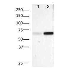
- Lysates prepared from HeLa cells that were untreated (lane 1) or treated with 10 uM TNF alpha for 10 min (lane 2) were run under reducing conditions and probed with 5 µg/mL of Anti-Human Phospho-NF kappa B p65 (S529) Purified, followed by Anti-Mouse IgG HRP.

Supportive validation
- Submitted by
- Invitrogen Antibodies (provider)
- Main image
- Experimental details
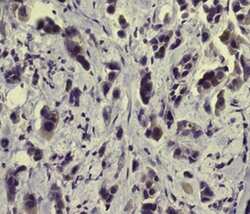
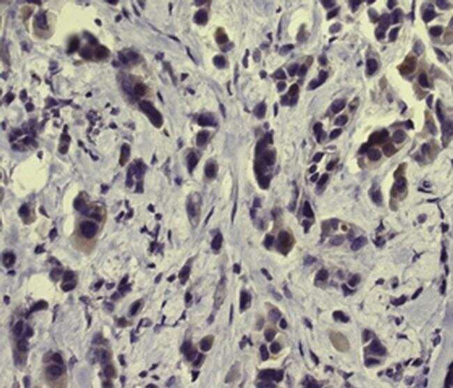
- Immunohistochemistry of formalin-fixed paraffin embedded human breast cancer tissue stained with 10 µg/mL of Anti-Human Phospho-NF kappa B p65 (S529) Purified followed by Anti-Mouse IgG Biotin, Streptavidin HRP, and visualized with DAB.Nuclei are counterstained with hematoxylin.
Supportive validation
- Submitted by
- Invitrogen Antibodies (provider)
- Main image
- Experimental details
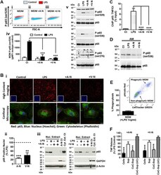
- Fig. 3 The impact of neutrophils on NF-kappaB activity in MDM. a LPS-induced (1 ng/ml; 20 min) p65 phosphorylation measured by (i-iv) flow cytometry (serine 529; n = 4) and (v) immunoblotting (serine 536, 529 and 276; n = 3) in MDM co-cultured with apoptotic or viable neutrophils (total time point: 30 min). b LPS-induced (1 ng/ml; 20 min) p65 nuclear translocation assessed by (i) quantitative high-content imaging and confocal microscopy (scale bars 100 mum and 7.5 mum, respectively), (ii) quantification of p65 nuclear translocation from high-content imaging ( n = 8) and (iii) immunoblotting in MDM co-cultured with apoptotic and viable neutrophils ( n = 3). Full images of individual stains and merged staining panels are available in supplemental data (supplemental data Fig. 1 ). c LPS-induced (1 ng/ml; 1 h) p65 binding to the TNF promotor in MDM co-cultured with apoptotic or viable neutrophils ( n = 3). d LPS-induced (1 ng/ml; 20 min) p65 phosphorylation in alveolar macrophages ( n = 3). e Phagocytosis of apoptotic neutrophils labelled with CellTracker Green and pHrodo Red by MDM at 40 min of co-culture in the presence of LPS (1 ng/ml; n = 14). f LPS-induced (1 ng/ml; 6 h) TNF release from MDM co-incubated with either apoptotic or viable neutrophils (cells), the supernatants from apoptotic or viable neutrophils cultured alone for 6 h (Sup) or the supernatants from apoptotic or viable neutrophils co-cultured with MDM for 6 h before (CoC-Sup) or after (CoC-Sup (UC) ultracentrifu
