 Explore
 Validate
 Learn
Learn Western blot
Antibody data
- Antibody Data
- Antigen structure
- References [3]
- Comments [0]
- Validations
- Western blot [1]
- Immunocytochemistry [1]
- Flow cytometry [1]
Submit
Validation data
Reference
Comment
Report error
- Product number
- 44-1170G - Provider product page

- Provider
- Invitrogen Antibodies
- Product name
- Phospho-4EBP1 (Thr46) Polyclonal Antibody
- Antibody type
- Polyclonal
- Antigen
- Synthetic peptide
- Reactivity
- Human, Mouse
- Host
- Rabbit
- Isotype
- IgG
- Vial size
- 100 µL
- Storage
- -20°C
Submitted references Impact of porcine reproductive and respiratory syndrome virus on muscle metabolism of growing pigs1.
Downregulated Translation Initiation Signaling Predisposes Low-Birth-Weight Neonatal Pigs to Slower Rates of Muscle Protein Synthesis.
Impact of prolonged leucine supplementation on protein synthesis and lean growth in neonatal pigs.
Helm ET, Curry SM, De Mille CM, Schweer WP, Burrough ER, Zuber EA, Lonergan SM, Gabler NK
Journal of animal science 2019 Jul 30;97(8):3213-3227
Journal of animal science 2019 Jul 30;97(8):3213-3227
Downregulated Translation Initiation Signaling Predisposes Low-Birth-Weight Neonatal Pigs to Slower Rates of Muscle Protein Synthesis.
Chen Y, McCauley SR, Johnson SE, Rhoads RP, El-Kadi SW
Frontiers in physiology 2017;8:482
Frontiers in physiology 2017;8:482
Impact of prolonged leucine supplementation on protein synthesis and lean growth in neonatal pigs.
Columbus DA, Steinhoff-Wagner J, Suryawan A, Nguyen HV, Hernandez-Garcia A, Fiorotto ML, Davis TA
American journal of physiology. Endocrinology and metabolism 2015 Sep 15;309(6):E601-10
American journal of physiology. Endocrinology and metabolism 2015 Sep 15;309(6):E601-10
No comments: Submit comment
Supportive validation
- Submitted by
- Invitrogen Antibodies (provider)
- Main image

- Experimental details
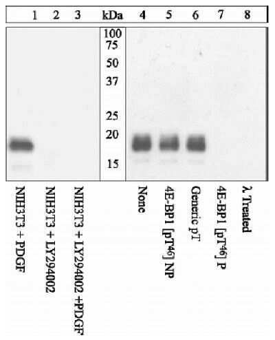
- Peptide Competition Lysates prepared from NIH3T3 cells left untreated or treated with PI-3K inhibitor LY2904002 (lanes 2 & 3), prior to PDGF stimulation (lanes 1 & 3) and from EGF-treated HEK293 cells (4-8), were incubated with 4E-BP1 (pT46) antib
Supportive validation
- Submitted by
- Invitrogen Antibodies (provider)
- Main image
- Experimental details
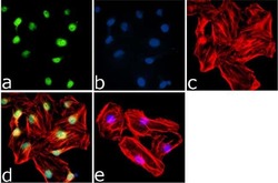
- Immunofluorescence analysis of 4EBP1 (pT46) was done on 70% confluent log phase HeLa cells. The cells were fixed with 4% paraformaldehyde for 15 minutes, permeabilized with 0.25% Triton™ X-100 for 10 minutes, and blocked with 5% BSA for 1 hour at room temperature. The cells were labeled with 4EBP1 (pT46) Rabbit Polyclonal Antibody (Product # 44-1170G) at 1:250 dilution in 1% BSA and incubated for 3 hours at room temperature and then labeled with Goat anti-Rabbit IgG (H+L) Superclonal™ Secondary Antibody, Alexa Fluor® 488 conjugate (Product # A27034) at a dilution of 1:2000 for 45 minutes at room temperature (Panel a: green). Nuclei (Panel b: blue) were stained with SlowFade® Gold Antifade Mountant with DAPI (Product # S36938). F-actin (Panel c: red) was stained with Rhodamine Phalloidin (Product # R415, 1:300). Panel d is a merged image showing Nuclear localization. Panel e is a no primary antibody control. The images were captured at 60X magnification.
Supportive validation
- Submitted by
- Invitrogen Antibodies (provider)
- Main image
- Experimental details
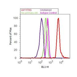
- Flow cytometry analysis of 4E-BP1 [pT46] was done on MCF7 cells treated with Insulin (100nM, 10 minutes). Cells were fixed with 70% ethanol for 10 minutes, permeabilized with 0.25% Triton™ X-100 for 20 minutes, and blocked with 5% BSA for 30 minutes at room temperature. Cells were labeled with 4E-BP1 [pT46] Rabbit Polyclonal Antibody (441170G, red histogram) or with rabbit isotype control (pink histogram) at 3-5 ug/million cells in 2.5% BSA. After incubation at room temperature for 2 hours, the cells were labeled with Alexa Fluor® 488 Goat Anti-Rabbit Secondary Antibody (A11008) at a dilution of 1:400 for 30 minutes at room temperature. The representative 10,000 cells were acquired and analyzed for each sample using an Attune® Acoustic Focusing Cytometer. The purple histogram represents unstained control cells and the green histogram represents no-primary-antibody control.
