Explore
 Validate
 Learn
Learn Western blot
Antibody data
- Antibody Data
- Antigen structure
- References [0]
- Comments [0]
- Validations
- Western blot [3]
- Immunocytochemistry [1]
- Immunohistochemistry [5]
Submit
Validation data
Reference
Comment
Report error
- Product number
- PA5-34759 - Provider product page

- Provider
- Invitrogen Antibodies
- Product name
- NF-H Polyclonal Antibody
- Antibody type
- Polyclonal
- Antigen
- Recombinant protein fragment
- Description
- Recommended positive controls: rat brain, IMR32.
- Concentration
- 0.71 mg/mL
No comments: Submit comment
Supportive validation
- Submitted by
- Invitrogen Antibodies (provider)
- Main image

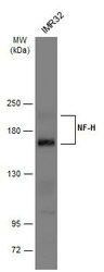
- Experimental details
- Western blot analysis of Neurofilament, Heavy chain using 30 µg of HeLa lysate. Samples were loaded onto a 7.5% SDS-PAGE gel and probed with a Neurofilament, Heavy chain polyclonal antibody (Product # PA5-34759) at a dilution of 1:1000.

- Submitted by
- Invitrogen Antibodies (provider)
- Main image
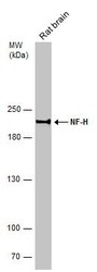
- Experimental details
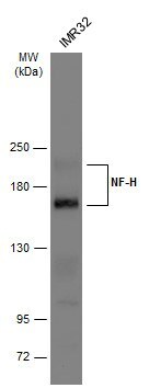
- Western Blot analysis of NF-H was performed by separating 50 µg of rat tissue extract by 5% SDS-PAGE. Proteins were transferred to a membrane and probed with a NF-H Polyclonal Antibody (Product # PA5-34759) at a dilution of 1:500.
- Submitted by
- Invitrogen Antibodies (provider)
- Main image
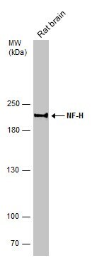
- Experimental details
- Western blot analysis of NF-H was performed by separating 30 µg of whole cell extract by 5% SDS-PAGE. Proteins were transferred to a membrane and probed with a NF-H Polyclonal Antibody (Product # PA5-34759) at a dilution of 1:500. The HRP-conjugated anti-rabbit IgG antibody was used to detect the primary antibody.
Supportive validation
- Submitted by
- Invitrogen Antibodies (provider)
- Main image
- Experimental details
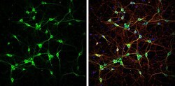
- Immunocytochemistry-Immunofluorescence analysis of NF-H was performed in DIV9 rat E18 primary cortical neurons fixed in 4% paraformaldehyde at RT for 15 min. Green: NF-H Polyclonal Antibody (Product # PA5-34759) diluted at 1:500. Red: beta Tubulin 3/ Tuj1, stained by beta Tubulin 3/ Tuj1 antibody. Blue: Fluoroshield with DAPI.

Supportive validation
- Submitted by
- Invitrogen Antibodies (provider)
- Main image

- Experimental details
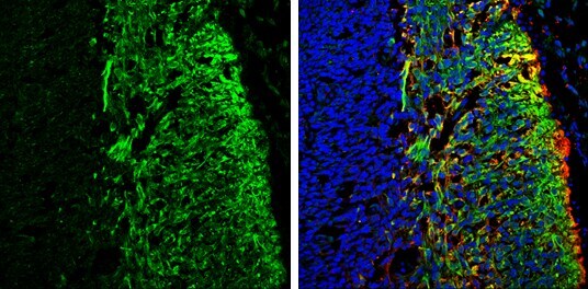
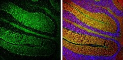
- Immunohistochemistry (Frozen) analysis of NF-H was performed in frozen sectioned E13.5 Rat brain tissue using NF-H Polyclonal Antibody (Product # PA5-34759) at a dilution of 1:250 (Green). Red: beta Tubulin 3/ TUJ1, a mature neuron marker, stained by beta Tubulin 3/ TUJ1 antibody diluted at 1:500. Blue: Fluoroshield with DAPI.
- Submitted by
- Invitrogen Antibodies (provider)
- Main image

- Experimental details
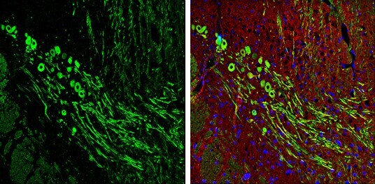
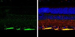
- Immunohistochemistry (Frozen) analysis of NF-H was performed in frozen-sectioned adult mouse cerebellum tissue using NF-H Polyclonal Antibody (Product # PA5-34759) at a dilution of 1:250 (Green). Red: beta Tubulin 3/ TUJ1, stained by beta Tubulin 3/ TUJ1 antibody diluted at 1:500. Blue: Fluoroshield with DAPI.
- Submitted by
- Invitrogen Antibodies (provider)
- Main image
- Experimental details
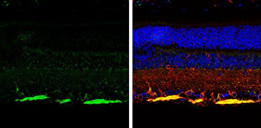
- Immunohistochemistry (Frozen) analysis of NF-H was performed in frozen-sectioned adult mouse cerebellum tissue using NF-H Polyclonal Antibody (Product # PA5-34759) at a dilution of 1:250 (Green). Red: beta Tubulin 3/ TUJ1, stained by beta Tubulin 3/ TUJ1 antibody diluted at 1:500. Blue: Fluoroshield with DAPI.

- Submitted by
- Invitrogen Antibodies (provider)
- Main image
- Experimental details
- Immunohistochemistry (Paraffin) analysis of NF-H was performed in paraffin-Embedded adult mouse retina tissue using Green: Vimentin Polyclonal Antibody (Product # PA5-27231) at a dilution of 1:250. Red: beta Tubulin 3/ TUJ1, stained by beta Tubulin 3/ TUJ1 antibody diluted at 1:500. Blue: Fluoroshield with DAPI.
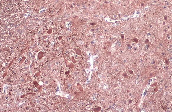
- Submitted by
- Invitrogen Antibodies (provider)
- Main image
- Experimental details
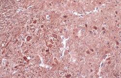
- NF-H Polyclonal Antibody detects NF-H protein at cytoplasm by immunohistochemical analysis. Sample: Paraffin-embedded mouse spinal cord. NF-H stained by NF-H Polyclonal Antibody (Product # PA5-34759) diluted at 1:500. Antigen Retrieval: Citrate buffer, pH 6.0, 15 min.