Explore
 Validate
 Learn
Learn Western blot
Antibody data
- Antibody Data
- Antigen structure
- References [14]
- Comments [0]
- Validations
- Western blot [2]
- Immunocytochemistry [2]
- Immunohistochemistry [3]
- Flow cytometry [1]
Submit
Validation data
Reference
Comment
Report error
- Product number
- MA5-12135 - Provider product page

- Provider
- Invitrogen Antibodies
- Product name
- Cytokeratin HMW Monoclonal Antibody (34BetaE12)
- Antibody type
- Monoclonal
- Antigen
- Other
- Description
- MA5-12135 targets Cytokeratin HMW and it recognizes keratin 1, 5, 10, and 14 in the Moll catalog (MW 68kDa, 58kDa, 56.5kDa, and 50kDa), respectively. The antibody has been tested in ICC/IF, WB and IHC (P) applications and shows reactivity with Equine, Human, Non-human primate, and Rabbit samples. This antibody does not react with rat tissue in Western blot applications.
- Antibody clone number
- 34BetaE12
- Concentration
- Conc. Not Determined
Submitted references Mechanisms of Endogenous HIV-1 Reactivation by Endocervical Epithelial Cells.
Reevaluation of MAML2 fusion-negative mucoepidermoid carcinoma: a subgroup being actually hyalinizing clear cell carcinoma of the salivary gland with EWSR1 translocation.
Sinonasal teratocarcinosarcoma: a clinical and pathological analysis.
Romanowsky-Giemsa as a counterstain for immunohistochemistry: optimizing a traditional reagent.
Malignant transformation of adenomyoepithelioma of the breast by a monophasic population: a report of two cases and review of literature.
A clinicopathological study of the significance of the proportion of choroid morphology in chordoid meningioma.
Chemical castration and anti-androgens induce differential gene expression in prostate cancer.
Identification of distinct gene expression profiles between esophageal squamous cell carcinoma and adjacent normal epithelial tissues.
The prognostic impact of O6-methylguanine DNA methyltransferase and epidermal growth factor receptor expressions on primary gliosarcoma: a clinicopathologic and immunohistochemical study of seven cases at a single institution.
Routine dual-color immunostaining with a 3-antibody cocktail improves the detection of small cancers in prostate needle biopsies.
Clear cell meningioma with frequent chordoid features and aggressive behavior: a clinicopathologic study of ten cases at a single institution.
Chordoid meningioma: a clinicopathologic study of 11 cases at a single institution.
Differences in the structural features of atypical adenomatous hyperplasia and low-grade prostatic adenocarcinoma.
Chronic inflammation in benign prostate hyperplasia is associated with focal upregulation of cyclooxygenase-2, Bcl-2, and cell proliferation in the glandular epithelium.
Gornalusse GG, Valdez R, Fenkart G, Vojtech L, Fleming LM, Pandey U, Hughes SM, Levy CN, Dela Cruz EJ, Calienes FL, Kirby AC, Fialkow MF, Lentz GM, Wagoner J, Jing L, Koelle DM, Polyak SJ, Fredricks DN, McElrath MJ, Wald A, Hladik F
Journal of virology 2020 Apr 16;94(9)
Journal of virology 2020 Apr 16;94(9)
Reevaluation of MAML2 fusion-negative mucoepidermoid carcinoma: a subgroup being actually hyalinizing clear cell carcinoma of the salivary gland with EWSR1 translocation.
Hsieh MS, Wang H, Lee YH, Ko JY, Chang YL
Human pathology 2017 Mar;61:9-18
Human pathology 2017 Mar;61:9-18
Sinonasal teratocarcinosarcoma: a clinical and pathological analysis.
Yang S, Sun R, Liang J, Zhou Z, Zhou J, Rui J
International journal of surgical pathology 2013 Feb;21(1):37-43
International journal of surgical pathology 2013 Feb;21(1):37-43
Romanowsky-Giemsa as a counterstain for immunohistochemistry: optimizing a traditional reagent.
Stefanović D, Stefanović M, Nikin Z
Biotechnic & histochemistry : official publication of the Biological Stain Commission 2013 Aug;88(6):329-35
Biotechnic & histochemistry : official publication of the Biological Stain Commission 2013 Aug;88(6):329-35
Malignant transformation of adenomyoepithelioma of the breast by a monophasic population: a report of two cases and review of literature.
Marian C, Boila A, Soanca D, Malau M, Podeanu DM, Resetkova E, Stolnicu S
APMIS : acta pathologica, microbiologica, et immunologica Scandinavica 2013 Apr;121(4):272-9
APMIS : acta pathologica, microbiologica, et immunologica Scandinavica 2013 Apr;121(4):272-9
A clinicopathological study of the significance of the proportion of choroid morphology in chordoid meningioma.
Lin JW, Lu CH, Lin WC, Wu YT, Huang YJ, Shih FY, Ho JT, Chuang MJ
Journal of clinical neuroscience : official journal of the Neurosurgical Society of Australasia 2012 Jun;19(6):836-43
Journal of clinical neuroscience : official journal of the Neurosurgical Society of Australasia 2012 Jun;19(6):836-43
Chemical castration and anti-androgens induce differential gene expression in prostate cancer.
Lehmusvaara S, Erkkilä T, Urbanucci A, Waltering K, Seppälä J, Larjo A, Tuominen VJ, Isola J, Kujala P, Lähdesmäki H, Kaipia A, Tammela TLj, Visakorpi T
The Journal of pathology 2012 Jul;227(3):336-45
The Journal of pathology 2012 Jul;227(3):336-45
Identification of distinct gene expression profiles between esophageal squamous cell carcinoma and adjacent normal epithelial tissues.
Tao Y, Chai D, Ma L, Zhang T, Feng Z, Cheng Z, Wu S, Qin Y, Lai M
The Tohoku journal of experimental medicine 2012 Apr;226(4):301-11
The Tohoku journal of experimental medicine 2012 Apr;226(4):301-11
The prognostic impact of O6-methylguanine DNA methyltransferase and epidermal growth factor receptor expressions on primary gliosarcoma: a clinicopathologic and immunohistochemical study of seven cases at a single institution.
Lin JW, Wu YT, Chang IW
Indian journal of pathology & microbiology 2011 Oct-Dec;54(4):683-7
Indian journal of pathology & microbiology 2011 Oct-Dec;54(4):683-7
Routine dual-color immunostaining with a 3-antibody cocktail improves the detection of small cancers in prostate needle biopsies.
Tolonen TT, Kujala PM, Laurila M, Tirkkonen M, Ilvesaro J, Tuominen VJ, Tammela TL, Isola J
Human pathology 2011 Nov;42(11):1635-42
Human pathology 2011 Nov;42(11):1635-42
Clear cell meningioma with frequent chordoid features and aggressive behavior: a clinicopathologic study of ten cases at a single institution.
Chen HK, Wu YT, Lin YJ, Lin JW
Journal of neuro-oncology 2011 Jul;103(3):551-9
Journal of neuro-oncology 2011 Jul;103(3):551-9
Chordoid meningioma: a clinicopathologic study of 11 cases at a single institution.
Lin JW, Ho JT, Lin YJ, Wu YT
Journal of neuro-oncology 2010 Dec;100(3):465-73
Journal of neuro-oncology 2010 Dec;100(3):465-73
Differences in the structural features of atypical adenomatous hyperplasia and low-grade prostatic adenocarcinoma.
Midi A, Tecimer T, Bozkurt S, Ozkan N
Indian journal of urology : IJU : journal of the Urological Society of India 2008 Apr;24(2):169-77
Indian journal of urology : IJU : journal of the Urological Society of India 2008 Apr;24(2):169-77
Chronic inflammation in benign prostate hyperplasia is associated with focal upregulation of cyclooxygenase-2, Bcl-2, and cell proliferation in the glandular epithelium.
Wang W, Bergh A, Damber JE
The Prostate 2004 Sep 15;61(1):60-72
The Prostate 2004 Sep 15;61(1):60-72
No comments: Submit comment
Supportive validation
- Submitted by
- Invitrogen Antibodies (provider)
- Main image

- Experimental details
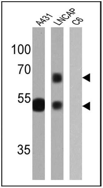
- Western blot analysis of Cytokeratin HMW was performed by loading 25 µg of A431 (lane 1), LNCAP (lane 2) and C6 (lane 3) cell lysates onto an SDS polyacrylamide gel. Proteins were transferred to a PVDF membrane and blocked at 4ºC overnight. The membrane was probed with a Cytokeratin HMW monoclonal antibody (Product # MA5-12135) at a dilution of 1:2000 overnight at 4°C, washed in TBST, and probed with an HRP-conjugated secondary antibody for 1 hr at room temperature in the dark. Chemiluminescent detection was performed using Pierce ECL Plus Western Blotting Substrate (Product # 32132). Results show a band at ~50-67 kDa in A431 and LNCAP cells.
- Submitted by
- Invitrogen Antibodies (provider)
- Main image
- Experimental details
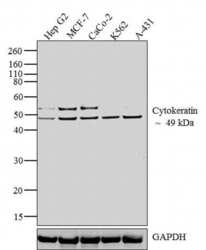
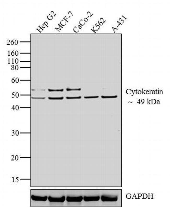
- Western blot analysis was performed on whole cell extracts (30 µg lysate) of Hep G2 (lane 1), MCF-7 (lane 2), CaCo-2 (lane 3), K562 (lane 4) and A-431 (lane 5). The blots were probed with Anti-Cytokeratin Mouse Monoclonal Antibody (Product # MA5-12135, 1:1000-1:10,000 dilution) and detected by chemiluminescence Goat anti-Mouse IgG (H+L) Secondary Antibody, HRP conjugate (Product # 62-6520, 1:4000 dilution). A 49 kDa band corresponding to Cytokeratin was observed across the cell lines tested along with an additional band at ~ 52 kDa in Hep G2, MCF-7 and CaCo-2. Known quantity of protein samples were electrophoresed using Novex® NuPAGE® 12 % Bis-Tris gel (Product # NP0342BOX), XCell SureLock™ Electrophoresis System (Product # EI0002) and Novex® Sharp Pre-Stained Protein Standard (Product # LC5800). Resolved proteins were then transferred onto a nitrocellulose membrane with iBlot® 2 Dry Blotting System (Product # IB21001). The membrane was probed with the relevant primary and secondary Antibody following blocking with 5 % skimmed milk. Chemiluminescent detection was performed using Pierce™ ECL Western Blotting Substrate (Product # 32106).
Supportive validation
- Submitted by
- Invitrogen Antibodies (provider)
- Main image
- Experimental details
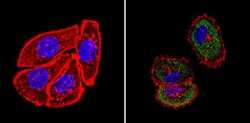
- Immunofluorescent analysis of Cytokeratin HMW (green) showing staining in the cytoplasm of A431 cells (right) compared to a negative control without primary antibody (left). Formalin-fixed cells were permeabilized with 0.1% Triton X-100 in TBS for 5-10 minutes and blocked with 3% BSA-PBS for 30 minutes at room temperature. Cells were probed with a Cytokeratin HMW monoclonal antibody (Product # MA5-12135) in 3% BSA-PBS at a dilution of 1:100 and incubated overnight at 4ºC in a humidified chamber. Cells were washed with PBST and incubated with a DyLight-conjugated secondary antibody in PBS at room temperature in the dark. F-actin (red) was stained with a fluorescent red phalloidin and nuclei (blue) were stained with Hoechst or DAPI. Images were taken at a magnification of 60x.
- Submitted by
- Invitrogen Antibodies (provider)
- Main image

- Experimental details
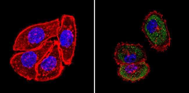
- Immunofluorescence analysis of Cytokeratin HMW was done on 70% confluent log phase A431 cells. The cells were fixed with 4% paraformaldehyde for 10 minutes, permeabilized with 0.1% Triton™ X-100 for 10 minutes, and blocked with 1% BSA for 1 hour at room temperature. The cells were labeled with of Cytokeratin HMW (34BetaE12) Mouse Monoclonal Antibody (Product # MA5-12135) at 1:250 dilution in0.1% BSA and incubated for 3 hours at room temperature and then labeled with Goat anti-Mouse IgG (H+L) Superclonal™ Secondary Antibody, Alexa Fluor® 488 conjugate (Product # A28175) at a dilution of 1:2000 for 45 minutes at room temperature (Panel a: green). Nuclei (Panel b: blue) were stained with SlowFade® Gold Antifade Mountant with DAPI (Product # S36938). F-actin (Panel c: red) was stained with Rhodamine Phalloidin (Product # R415, 1:300). Panel d is a merged image showing cytoplasmic localization. Panel e is a no primary antibody control. The images were captured at 60X magnification.

Supportive validation
- Submitted by
- Invitrogen Antibodies (provider)
- Main image
- Experimental details
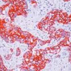
- Formalin-fixed, paraffin-embedded human breast cancer stained with Cytokeratin HMW antibody (Product # MA5-12135) using peroxidase-conjugate and AEC chromogen. Note cytoplasmic staining of tumor cells.
- Submitted by
- Invitrogen Antibodies (provider)
- Main image
- Experimental details
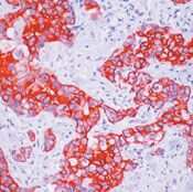
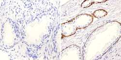
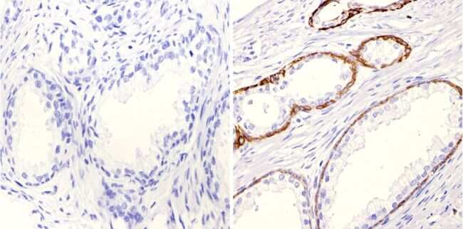
- Immunohistochemistry analysis of Cytokeratin HMW showing positive staining in the cytoplasm of paraffin-treated Human prostate tissue (right) compared with a negative control in the absence of primary antibody (left). To expose target proteins, antigen retrieval method was performed using 10mM sodium citrate (pH 6.0), microwaved for 8-15 min. Following antigen retrieval, tissues were blocked in 3% H2O2-methanol for 15 min at room temperature, washed with ddH2O and PBS, and then probed with a Cytokeratin HMW monoclonal antibody (Product # MA5-12135) diluted by 3% BSA-PBS at a dilution of 1:100 overnight at 4°C in a humidified chamber. Tissues were washed extensively PBST and detection was performed using an HRP-conjugated secondary antibody followed by colorimetric detection using a DAB kit. Tissues were counterstained with hematoxylin and dehydrated with ethanol and xylene to prep for mounting.
- Submitted by
- Invitrogen Antibodies (provider)
- Main image

- Experimental details
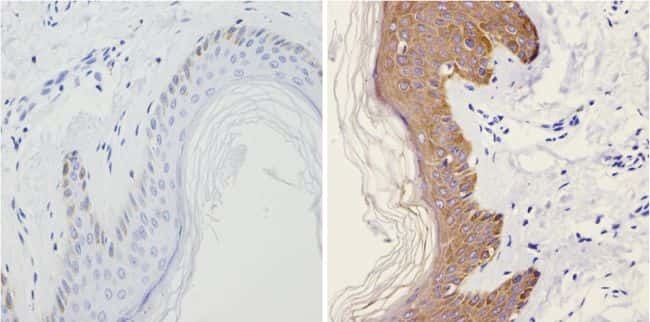
- Immunohistochemistry analysis of Cytokeratin HMW showing positive staining in the cytoplasm of paraffin-treated Human skin tissue (right) compared with a negative control in the absence of primary antibody (left). To expose target proteins, antigen retrieval method was performed using 10mM sodium citrate (pH 6.0), microwaved for 8-15 min. Following antigen retrieval, tissues were blocked in 3% H2O2-methanol for 15 min at room temperature, washed with ddH2O and PBS, and then probed with a Cytokeratin HMW monoclonal antibody (Product # MA5-12135) diluted by 3% BSA-PBS at a dilution of 1:200 overnight at 4°C in a humidified chamber. Tissues were washed extensively PBST and detection was performed using an HRP-conjugated secondary antibody followed by colorimetric detection using a DAB kit. Tissues were counterstained with hematoxylin and dehydrated with ethanol and xylene to prep for mounting.
Supportive validation
- Submitted by
- Invitrogen Antibodies (provider)
- Main image
- Experimental details
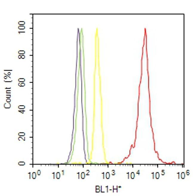
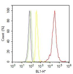
- Flow cytometry analysis of Cytokeratin HMW was done on A-431 cells. Cells were fixed with 70% ethanol for 10 minutes, permeabilized with 0.25% Triton™ X-100 for 20 minutes, and blocked with 5% BSA for 30 minutes at room temperature. Cells were labeled with Cytokeratin HMW Mouse Monoclonal Antibody (MA512135, red histogram) or with mouse isotype control (yellow histogram) at 3-5 ug/million cells in 2.5% BSA. After incubation at room temperature for 2 hours, the cells were labeled with Alexa Fluor® 488 Rabbit Anti-Mouse Secondary Antibody (A11059) at a dilution of 1:400 for 30 minutes at room temperature. The representative 10,000 cells were acquired and analyzed for each sample using an Attune® Acoustic Focusing Cytometer. The purple histogram represents unstained control cells and the green histogram represents no-primary-antibody control.