Explore
 Validate
 Learn
LearnMA5-17054
antibody from Invitrogen Antibodies
Targeting: CDKN2A
ARF, CDK4I, CDKN2, CMM2, INK4, INK4a, MLM, MTS1, p14, p14ARF, p16, p16INK4a, p19, p19Arf
 Western blot
Antibody data
- Antibody Data
- Antigen structure
- References [0]
- Comments [0]
- Validations
- Western blot [6]
- Immunocytochemistry [1]
- Immunohistochemistry [2]
- Flow cytometry [1]
Submit
Validation data
Reference
Comment
Report error
- Product number
- MA5-17054 - Provider product page

- Provider
- Invitrogen Antibodies
- Product name
- p16INK4a Monoclonal Antibody (1D7D2)
- Antibody type
- Monoclonal
- Antigen
- Purifed from natural sources
- Description
- MA5-17054 targets CDKN2A in FACS, IHC, pep-ELISA, and WB applications and shows reactivity with Human samples.
- Antibody clone number
- 1D7D2
- Concentration
- Conc. Not Determined
No comments: Submit comment
Supportive validation
- Submitted by
- Invitrogen Antibodies (provider)
- Main image
- Experimental details
- Western blot analysis of CDKN2A using a CDKN2A monoclonal antibody (Product # MA5-17054) against a human CDKN2A recombinant protein.

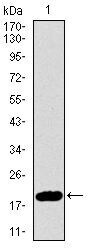
- Submitted by
- Invitrogen Antibodies (provider)
- Main image

- Experimental details
- Western blot analysis of CDKN2A using CDKN2A monoclonal antibody (Product # MA5-17054) in HEK293 (1) and CDKN2A (AA: 1-156) human IgG Fc transfected HEK293 (2) cell lysate.
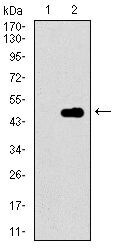
- Submitted by
- Invitrogen Antibodies (provider)
- Main image
- Experimental details
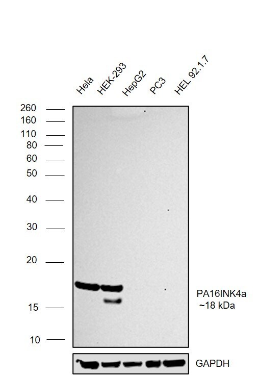
- Western blot was performed using Anti-p16INK4a Monoclonal Antibody (1D7D2) (Product # MA5-17054) and a ~18 kDa band corresponding to CDKN2A was observed across cell lines tested. Whole cell extracts (30 µg lysate) of HeLa (Lane 1), HEK-293 (Lane 2), Hep G2 (Lane 3), PC-3 (Lane 4), HEL 92.1.7 (Lane 5) were electrophoresed using NuPAGE™ 12% Bis-Tris Protein Gel (Product # NP0341BOX). Resolved proteins were then transferred onto a nitrocellulose membrane (Product # IB23001) by iBlot® 2 Dry Blotting System (Product # IB21001). The blot was probed with the primary antibody (1:1000) and detected by chemiluminescence with Goat anti-Mouse IgG (H+L) Superclonal™ Recombinant Secondary Antibody, HRP (Product # A28177,1:20000) using the iBright™ FL1500 Imaging System (Product # A44115). Chemiluminescent detection was performed using SuperSignal™ West Pico PLUS Chemiluminescent Substrate (Product # 34580).
- Submitted by
- Invitrogen Antibodies (provider)
- Main image
- Experimental details
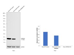
- Knockout of CDKN2A was achieved by CRISPR-Cas9 genome editing using LentiArray™ Lentiviral sgRNA (Product # A32042) (Assay ID CRISPR697306_LV) and LentiArray Cas9 Lentivirus (Product # A32064). Western blot analysis of CDKN2A was performed by loading 30 µg of HeLa wild type (Lane 1), HeLa CAS9 (Lane 2), HeLa CDKN2A KO (Lane 3) whole cell extracts. The blot was probed with Anti-p16INK4a Monoclonal Antibody (1D7D2)(Product # MA5-17054) using 1:2000 dilution and Goat anti-Mouse IgG (H+L), Superclonal™ Recombinant Secondary Antibody, HRP (Product # A28177). Loss of signal upon CRISPR mediated knockout (KO) using the LentiArray™ CRISPR product line confirms that antibody is specific to CDKN2A.
- Submitted by
- Invitrogen Antibodies (provider)
- Main image

- Experimental details
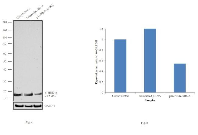
- Knockdown of p16INK4a was achieved by transfecting Caco-2 with p16INK4a specific siRNAs (Silencer® select Product # s216). Western blot analysis (Fig. a) was performed using whole cell extracts from the p16INK4a knockdown cells (lane 3), non-specific scrambled siRNA transfected cells (lane 2) and untransfected cells (lane 1). The blots were probed with p16INK4a Monoclonal Antibody (1D7D2) (Product # MA5-17054, 1:2000 dilution) and Goat anti-Mouse IgG (H+L) Superclonal™ Secondary Antibody, HRP conjugate (Product # A28177, 0.25 µg/ml 1:4000 dilution). Densitometric analysis of this western blot is shown in histogram (Fig. b). Decrease in signal upon siRNA mediated knock down confirms that antibody is specific to p16INK4a.
- Submitted by
- Invitrogen Antibodies (provider)
- Main image

- Experimental details
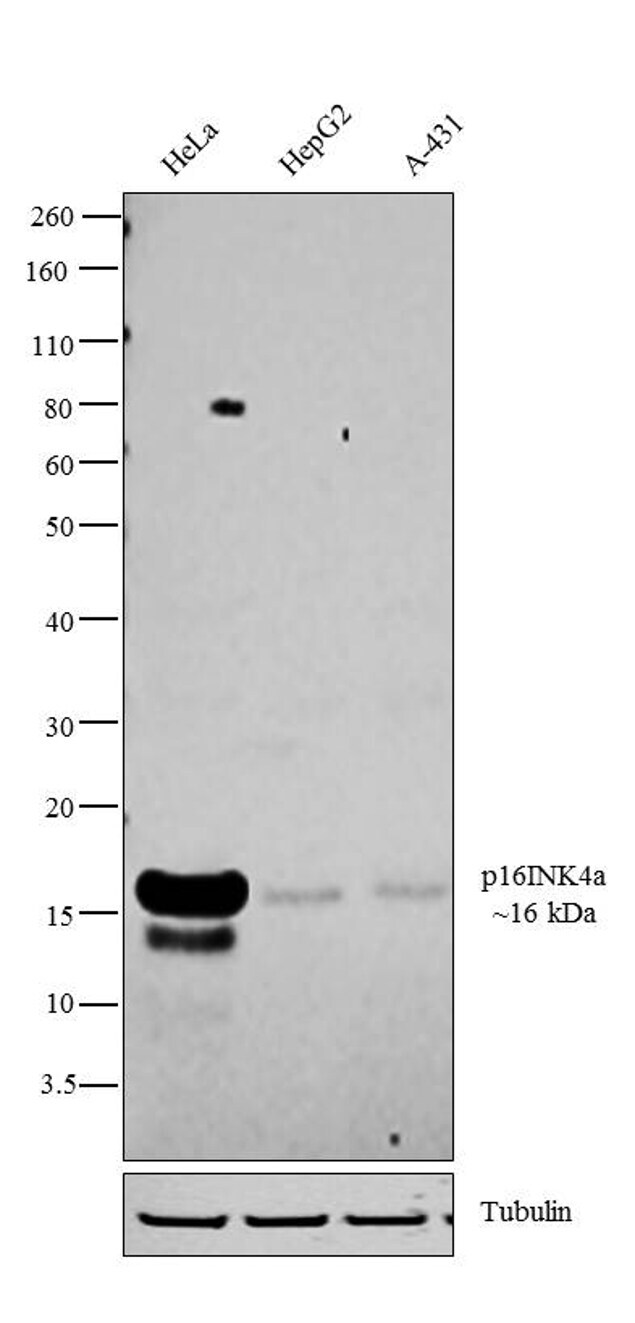
- Western blot analysis was performed on modified whole cell extracts (1% SDS) (30 µg lysate) of HeLa (Lane 1), Hep G2 (Lane 2) and A-431 (Lane 3). The blot was probed with Anti- p16INK4a Monoclonal Antibody (Product # MA5-17054, 1:1000 dilution) and detected by chemiluminescence using Goat anti-Mouse IgG (H+L) Superclonal™ Secondary Antibody, HRP conjugate (Product # A28177, 0.25 µg/ml, 1:4000 dilution). A 16 kDa band corresponding to p16INK4a was observed across the cell lines tested.
Supportive validation
- Submitted by
- Invitrogen Antibodies (provider)
- Main image
- Experimental details
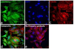
- Immunofluorescence analysis of CDKN2A was performed using 70% confluent log phase HeLa cells. The cells were fixed with 4% paraformaldehyde for 10 minutes, permeabilized with 0.1% Triton™ X-100 for 15 minutes, and blocked with 2% BSA for 45 minutes at room temperature. The cells were labeled with p16INK4a Monoclonal Antibody (1D7D2) (Product # MA5-17054) at 1:100 in 0.1% BSA, incubated at 4 degree celsius overnight and then labeled with Donkey anti-Mouse IgG (H+L) Highly Cross-Adsorbed Secondary Antibody, Alexa Fluor Plus 488 (Product # A32766), (1:2000), for 45 minutes at room temperature (Panel a: Green). Nuclei (Panel b:Blue) were stained with ProLong™ Diamond Antifade Mountant with DAPI (Product # P36962). F-actin (Panel c: Red) was stained with Rhodamine Phalloidin (Product # R415, 1:300). Panel d represents the merged image showing nucleus and cytoplasm localization. Panel e represents control cells with no primary antibody to assess background. The images were captured at 40X. magnification.
Supportive validation
- Submitted by
- Invitrogen Antibodies (provider)
- Main image
- Experimental details
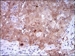
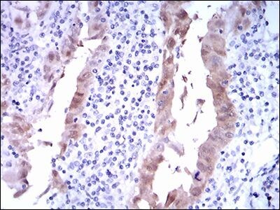
- Immunohistochemical analysis of paraffin-embedded lung cancer tissues using CDKN2A monoclonal antibody (Product # MA5-17054) followed with DAB staining.

- Submitted by
- Invitrogen Antibodies (provider)
- Main image

- Experimental details
- Immunohistochemical analysis of paraffin-embedded endometrial cancer tissues using CDKN2A monoclonal antibody (Product # MA5-17054) followed with DAB staining.
Supportive validation
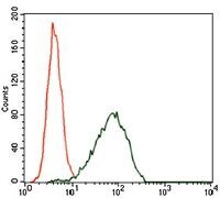
- Submitted by
- Invitrogen Antibodies (provider)
- Main image
- Experimental details
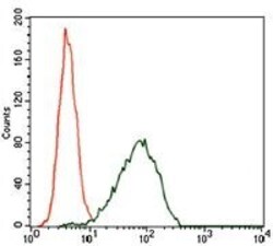
- Flow cytometric analysis of HEK293 cells using CDKN2A monoclonal antibody (Product # MA5-17054) (green) and negative control (red).