Explore
 Validate
 Learn
Learn Western blot
 Flow cytometry
Antibody data
- Antibody Data
- Antigen structure
- References [1]
- Comments [0]
- Validations
- Western blot [1]
- Immunohistochemistry [1]
- Other assay [1]
Submit
Validation data
Reference
Comment
Report error
- Product number
- MA5-16351 - Provider product page

- Provider
- Invitrogen Antibodies
- Product name
- Claudin 1 Monoclonal Antibody (SP128)
- Antibody type
- Monoclonal
- Antigen
- Synthetic peptide
- Description
- Heat-mediated antigen retrieval is recommended prior to staining, using a 1mM EDTA buffer, pH 8.0, for 10 minutes followed by cooling at room temperature for 20 min. Following antigen retrieval, incubate samples with primary antibody for 30 min at room temperature. A suggested positive control is liver, hepatocellular carcinoma or ovarian carcinoma.
- Reactivity
- Human
- Host
- Rabbit
- Isotype
- IgG
- Antibody clone number
- SP128
- Vial size
- 500 µL
- Storage
- Store at 4°C short term. For long term storage, store at -20°C, avoiding freeze/thaw cycles.
Submitted references Development of an Improved 3D in vitro Intestinal Model to Perform Permeability Studies of Paracellular Compounds.
Macedo MH, Martínez E, Barrias CC, Sarmento B
Frontiers in bioengineering and biotechnology 2020;8:524018
Frontiers in bioengineering and biotechnology 2020;8:524018
No comments: Submit comment
Supportive validation
- Submitted by
- Invitrogen Antibodies (provider)
- Main image
- Experimental details
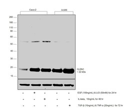
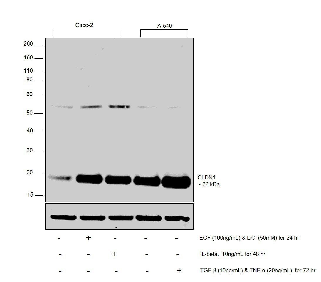
- Western blot was performed using Anti-CLDN1 Monoclonal Antibody (SP128) (Product # MA5-16351) and a 22 kDa band corresponding to CLDN1 was observed across cell lines tested and increased upon treatment. Membrane enriched extracts (30 µg lysate) of Caco-2 (Lane 1), Caco-2 treated with EGF (100ng/mL) and LiCl (50mM) simultaneously for 24 hr (Lane 2), Caco-2 treated with IL-beta (10ng/mL for 48 hr) (Lane 3), (25 ug lysate) of A-549 (Lane 4) and A-549 treated with TGF-beta (10ng/mL) and TNF-alpha (20ng/mL) simultaneously for 72 hr (Lane 5) were electrophoresed using Novex® NuPAGE® 12 % Bis-Tris gel (Product # NP0342BOX). Resolved proteins were then transferred onto a nitrocellulose membrane (Product # IB23001) by iBlot® 2 Dry Blotting System (Product # IB21001). The blot was probed with the primary antibody (1:1000 dilution) and detected by chemiluminescence with Goat anti-Rabbit IgG (H+L), Superclonal™ Recombinant Secondary Antibody, HRP (Product # A27036, 1:4000 dilution) using the iBright FL 1000 (Product # A32752). Chemiluminescent detection was performed using Novex® ECL Chemiluminescent Substrate Reagent Kit (Product # WP20005).
Supportive validation
- Submitted by
- Invitrogen Antibodies (provider)
- Main image
- Experimental details
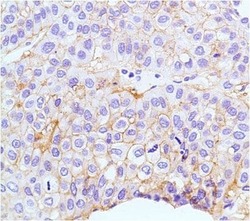
- Immunohistochemical analysis of Claudin-1 using anti-Claudin-1 Monoclonal Antibody (Product # MA5-16351) in Hepatocellular Carcinoma Cancer Tissue. The recommened dilution for this antibody in immunohistochemistry applications is 1:200.

Supportive validation
- Submitted by
- Invitrogen Antibodies (provider)
- Main image
- Experimental details
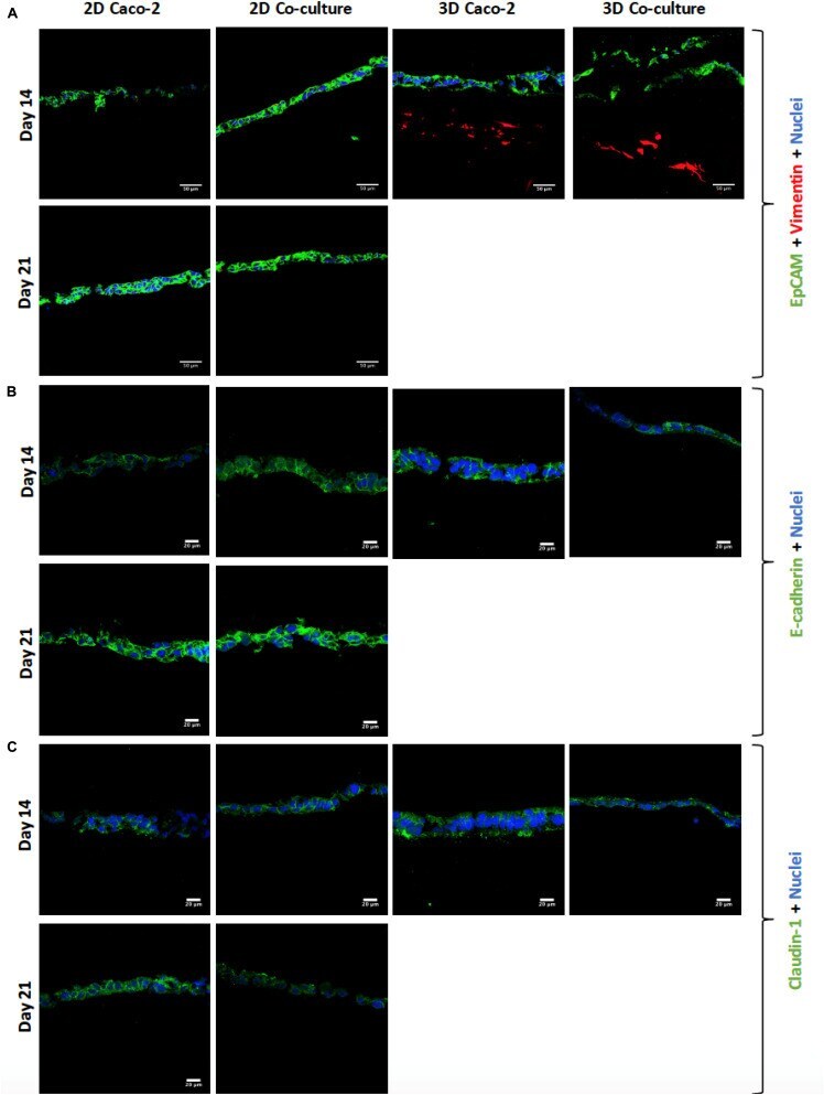
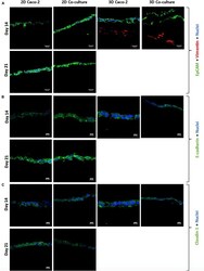
- FIGURE 6 (A) Visualization of cell layer formation by EpCAM, confirming the presence of a cell layer in all models. (B) Assessment and comparison of cellular adherens junctions in different models at different time points using E -cadherin. (C) Assessment and comparison of the tight junctions' protein claudin-1 in the different models at different time points. EpCAM, E -cadherin, and claudin-1 were labeled with Alexa-Fluor 488 (green), and nuclei were stained with 4',6-diamidino-2-phenylindole (DAPI) (blue).