Explore
 Validate
 Learn
Learn Western blot
 ELISA
ELISAAntibody data
- Antibody Data
- Antigen structure
- References [9]
- Comments [0]
- Validations
- Western blot [7]
- Flow cytometry [2]
Submit
Validation data
Reference
Comment
Report error
- Product number
- NBP1-47423 - Provider product page

- Provider
- Novus Biologicals
- Proper citation
- Novus Cat#NBP1-47423, RRID:AB_10010376
- Product name
- Mouse Monoclonal beta-Actin Antibody
- Antibody type
- Monoclonal
- Description
- Ammonium sulfate precipitation.
- Reactivity
- Human, Mouse, Rat, Simian
- Host
- Mouse
- Isotype
- IgG
- Vial size
- 0.1 ml
- Concentration
- 1.0 mg/ml
- Storage
- Store at 4C short term. Aliquot and store at -20C long term. Avoid freeze-thaw cycles.
Submitted references Biological activity and apoptotic signaling pathway of C11-functionalized cephalostatin 1 analogues.
Activation of Phospholipase C β by Gβγ and Gαq Involves C-Terminal Rearrangement to Release Autoinhibition.
Repurposing pyridoxamine for therapeutic intervention of intravascular cell-cell interactions in mouse models of sickle cell disease.
Mechanistic Study of TTF-1 Modulation of Cellular Sensitivity to Cisplatin.
Sterically stabilized siRNA:gold nanocomplexes enhance c-MYC silencing in a breast cancer cell model.
Consumption of Terpenoids-Rich Padina pavonia Extract Attenuates Hyperglycemia, Insulin Resistance and Oxidative Stress, and Upregulates PPARγ in a Rat Model of Type 2 Diabetes.
Cephalostatin 1 analogues activate apoptosis via the endoplasmic reticulum stress signaling pathway.
LRRK2 G2019S-induced mitochondrial DNA damage is LRRK2 kinase dependent and inhibition restores mtDNA integrity in Parkinson's disease.
DREAM plays an important role in platelet activation and thrombogenesis.
Nawasreh MM, Alzyoud EI, Al-Mazaydeh ZA, Rammaha MS, Yasin SR, Tahtamouni LH
Steroids 2020 Jun;158:108602
Steroids 2020 Jun;158:108602
Activation of Phospholipase C β by Gβγ and Gαq Involves C-Terminal Rearrangement to Release Autoinhibition.
Fisher IJ, Jenkins ML, Tall GG, Burke JE, Smrcka AV
Structure (London, England : 1993) 2020 Jul 7;28(7):810-819.e5
Structure (London, England : 1993) 2020 Jul 7;28(7):810-819.e5
Repurposing pyridoxamine for therapeutic intervention of intravascular cell-cell interactions in mouse models of sickle cell disease.
Li J, Jeong SY, Xiong B, Tseng A, Mahon AB, Isaacman S, Gordeuk VR, Cho J
Haematologica 2019 Oct 31;
Haematologica 2019 Oct 31;
Mechanistic Study of TTF-1 Modulation of Cellular Sensitivity to Cisplatin.
Phelps CA, Lindsey-Boltz L, Sancar A, Mu D
Scientific reports 2019 May 29;9(1):7990
Scientific reports 2019 May 29;9(1):7990
Sterically stabilized siRNA:gold nanocomplexes enhance c-MYC silencing in a breast cancer cell model.
Daniels AN, Singh M
Nanomedicine (London, England) 2019 Jun;14(11):1387-1401
Nanomedicine (London, England) 2019 Jun;14(11):1387-1401
Consumption of Terpenoids-Rich Padina pavonia Extract Attenuates Hyperglycemia, Insulin Resistance and Oxidative Stress, and Upregulates PPARγ in a Rat Model of Type 2 Diabetes.
Germoush MO, Elgebaly HA, Hassan S, Kamel EM, Bin-Jumah M, Mahmoud AM
Antioxidants (Basel, Switzerland) 2019 Dec 26;9(1)
Antioxidants (Basel, Switzerland) 2019 Dec 26;9(1)
Cephalostatin 1 analogues activate apoptosis via the endoplasmic reticulum stress signaling pathway.
Tahtamouni LH, Nawasreh MM, Al-Mazaydeh ZA, Al-Khateeb RA, Abdellatif RN, Bawadi RM, Bamburg JR, Yasin SR
European journal of pharmacology 2018 Jan 5;818:400-409
European journal of pharmacology 2018 Jan 5;818:400-409
LRRK2 G2019S-induced mitochondrial DNA damage is LRRK2 kinase dependent and inhibition restores mtDNA integrity in Parkinson's disease.
Howlett EH, Jensen N, Belmonte F, Zafar F, Hu X, Kluss J, Schüle B, Kaufman BA, Greenamyre JT, Sanders LH
Human molecular genetics 2017 Nov 15;26(22):4340-4351
Human molecular genetics 2017 Nov 15;26(22):4340-4351
DREAM plays an important role in platelet activation and thrombogenesis.
Kim K, Tseng A, Barazia A, Italiano JE, Cho J
Blood 2017 Jan 12;129(2):209-225
Blood 2017 Jan 12;129(2):209-225
No comments: Submit comment
Supportive validation
- Submitted by
- Novus Biologicals (provider)
- Main image

- Experimental details
- Simple Western: beta-Actin Antibody (8H10D10) [NBP1-47423] - Image shows a specific band for Beta Actin in 1.0 mg/mL of HeLa lysate. This experiment was performed under reducing conditions using the 12-230 kDa separation system.

- Submitted by
- Novus Biologicals (provider)
- Main image

- Experimental details
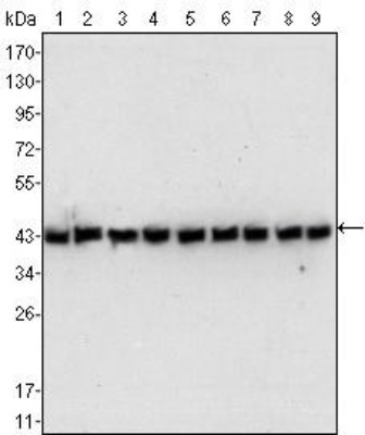
- Western Blot: beta-Actin Antibody (8H10D10) [NBP1-47423] - Analysis using anti-Beta Actin mAb against human, mouse, hamster, rat, and primate cell lystate(s): (1) NIH/3T3, (2) Jurkat, (3) HeLa, (4) CHO, (5) PC12, (6) HEK293, (7) COS, (8) A549, and (9) MCF-7. A specific band can be observed at a molecular weight of approximately 43 kDa in all lanes.
- Submitted by
- Novus Biologicals (provider)
- Main image
- Experimental details
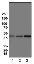
- Western Blot: beta-Actin Antibody (8H10D10) [NBP1-47423] - Analysis of Beta Actin expression in 1) HeLa 2) HepG2 and 3) Cos7 whole cell lysates.

- Submitted by
- Novus Biologicals (provider)
- Main image
- Experimental details
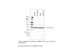
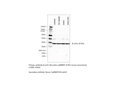
- Western Blot: beta-Actin Antibody (8H10D10) [NBP1-47423] - Western blot of pancreatic cell lines (CAPAN2, PATU8988T, DANG, and PANC1005). Primary antibody: beta-Actin (8H10D10), NBP1-47423, 1:1000 dilution. Secondary antibody: HRP conjugated Mouse IgG (H+L), NB7539. A specific band can be observed at a molecular weight of approximately 42 kDa. Image from verified customer review.
- Submitted by
- Novus Biologicals (provider)
- Main image

- Experimental details
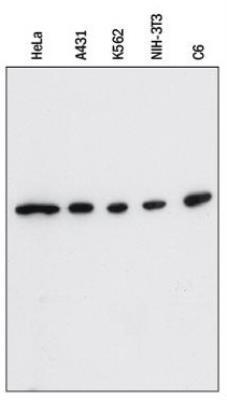
- Western Blot: beta-Actin Antibody (8H10D10) [NBP1-47423] - Lysates of HeLa human cervical epithelial carcinoma cell line, A431 human epithelial carcinoma cell line, K562 human chronic myelogenous leukemia cell line, NIH-3T3 mouse embryonic fibroblast cell line, and C6 rat glioma cell line were probed with 1:2500 mouse anti-beta-Actin monoclonal (NBP1-47423) followed by 1:2000 dilution of donkey anti-mouse IgG-HRP secondary antibody (HAF018).
- Submitted by
- Novus Biologicals (provider)
- Main image
- Experimental details
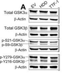
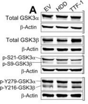
- Western Blot: beta-Actin Antibody (8H10D10) [NBP1-47423] - TTF-1 alters the phosphorylation of GSK3alpha/beta. (A) Panels of immunoblot images examining total GSK3alpha/beta and phosphorylation at specific Ser or Tyr residues in A549 transfectant cells. Beta-Actin is included as a loading control. Image collected and cropped by CiteAb from the following publication (http://www.nature.com/articles/s41598-019-44549-w), licensed under a CC-BY licence.
- Submitted by
- Novus Biologicals (provider)
- Main image

- Experimental details
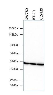
- Western Blot: beta-Actin Antibody (8H10D10) [NBP1-47423] - Three different human cancer cell lines (SW780, BT-20, and CLS439) were probed with the antibody. Image from verified customer review.
Supportive validation
- Submitted by
- Novus Biologicals (provider)
- Main image

- Experimental details
- Flow Cytometry: beta-Actin Antibody (8H10D10) [NBP1-47423] - Analysis of MCF-7 cells using anti-Beta Actin mAb (right) and negative control (left).

- Submitted by
- Novus Biologicals (provider)
- Main image
- Experimental details
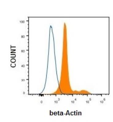
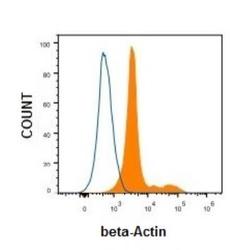
- Flow Cytometry: beta-Actin Antibody (8H10D10) [NBP1-47423] - Analysis of HeLa cells using mouse Monoclonal beta-Actin antibody (Orange) and Isotype control Antibody (Blue).