Explore
 Validate
 Learn
Learn Flow cytometry
Antibody data
- Antibody Data
- Antigen structure
- References [4]
- Comments [0]
- Validations
- Flow cytometry [1]
- Other assay [2]
Submit
Validation data
Reference
Comment
Report error
- Product number
- 25-0106-42 - Provider product page

- Provider
- Invitrogen Antibodies
- Product name
- CD10 Monoclonal Antibody (eBioCB-CALLA (CB-CALLA)), PE-Cyanine7, eBioscience™
- Antibody type
- Monoclonal
- Antigen
- Other
- Description
- Description: The eBioCB-CALLA monoclonal antibody recognizes human CD10 (CALLA, NEP, enkephalinase, Neprilysin), which is a 100 kDa, type II cell surface glycoprotein originally identified for its expression on most acute lymphoblastic leukemias (ALL). Subsequently, CD10 was shown to be the same molecule as the neutral endopeptidase (NEP), or KII-NA. CD10 is a Zn2+-dependent metallo-peptidase with endothelin, glucagon, gastrin, neurotensin and bradykinin included among its substrates. CD10 is involved in the regulation of chemotactic and inflammatory processes involving neutrophils. In B cells, CD10 regulates stromal cell-dependent B lymphopoiesis and expression has also been reported on mature B cells in germinal centres. In addition to the hematopoietic compartment, other major sites of CD10 expression are the brush border of enterocytes and renal tubules and glomeruli. There is partial blocking of the eBioCB-CALLA and MEM-78 monoclonal antibodies indicating that they recognize similar epitopes.
- Antibody clone number
- eBioCB-CALLA (CB-CALLA)
- Concentration
- 5 µL/Test
Submitted references Heterogeneous disease-propagating stem cells in juvenile myelomonocytic leukemia.
Elevated Calprotectin and Abnormal Myeloid Cell Subsets Discriminate Severe from Mild COVID-19.
HIV Malaria Co-Infection Is Associated with Atypical Memory B Cell Expansion and a Reduced Antibody Response to a Broad Array of Plasmodium falciparum Antigens in Rwandan Adults.
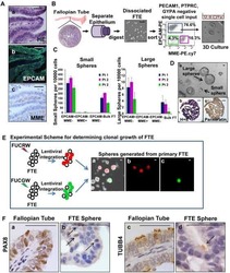
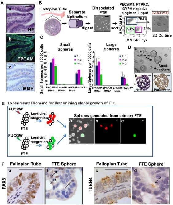
Stem-like epithelial cells are concentrated in the distal end of the fallopian tube: a site for injury and serous cancer initiation.
Louka E, Povinelli B, Rodriguez-Meira A, Buck G, Wen WX, Wang G, Sousos N, Ashley N, Hamblin A, Booth CAG, Roy A, Elliott N, Iskander D, de la Fuente J, Fordham N, O'Byrne S, Inglott S, Norfo R, Salio M, Thongjuea S, Rao A, Roberts I, Mead AJ
The Journal of experimental medicine 2021 Feb 1;218(2)
The Journal of experimental medicine 2021 Feb 1;218(2)
Elevated Calprotectin and Abnormal Myeloid Cell Subsets Discriminate Severe from Mild COVID-19.
Silvin A, Chapuis N, Dunsmore G, Goubet AG, Dubuisson A, Derosa L, Almire C, Hénon C, Kosmider O, Droin N, Rameau P, Catelain C, Alfaro A, Dussiau C, Friedrich C, Sourdeau E, Marin N, Szwebel TA, Cantin D, Mouthon L, Borderie D, Deloger M, Bredel D, Mouraud S, Drubay D, Andrieu M, Lhonneur AS, Saada V, Stoclin A, Willekens C, Pommeret F, Griscelli F, Ng LG, Zhang Z, Bost P, Amit I, Barlesi F, Marabelle A, Pène F, Gachot B, André F, Zitvogel L, Ginhoux F, Fontenay M, Solary E
Cell 2020 Sep 17;182(6):1401-1418.e18
Cell 2020 Sep 17;182(6):1401-1418.e18
HIV Malaria Co-Infection Is Associated with Atypical Memory B Cell Expansion and a Reduced Antibody Response to a Broad Array of Plasmodium falciparum Antigens in Rwandan Adults.
Subramaniam KS, Skinner J, Ivan E, Mutimura E, Kim RS, Feintuch CM, Portugal S, Anastos K, Crompton PD, Daily JP
PloS one 2015;10(4):e0124412
PloS one 2015;10(4):e0124412
Stem-like epithelial cells are concentrated in the distal end of the fallopian tube: a site for injury and serous cancer initiation.
Paik DY, Janzen DM, Schafenacker AM, Velasco VS, Shung MS, Cheng D, Huang J, Witte ON, Memarzadeh S
Stem cells (Dayton, Ohio) 2012 Nov;30(11):2487-97
Stem cells (Dayton, Ohio) 2012 Nov;30(11):2487-97
No comments: Submit comment
Supportive validation
- Submitted by
- Invitrogen Antibodies (provider)
- Main image
- Experimental details
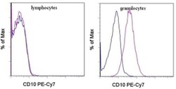
- Staining of normal human peripheral blood cells with Mouse IgG2b K Isotype Control PE-Cyanine7 (Product # 25-4732-81) (blue histogram) or Anti-Human CD10 PE-Cyanine7 (purple histogram). Cells in the lymphocyte gate (left) or granulocyte gate (right) were used for analysis.

Supportive validation
- Submitted by
- Invitrogen Antibodies (provider)
- Main image
- Experimental details
- NULL
- Submitted by
- Invitrogen Antibodies (provider)
- Main image
- Experimental details
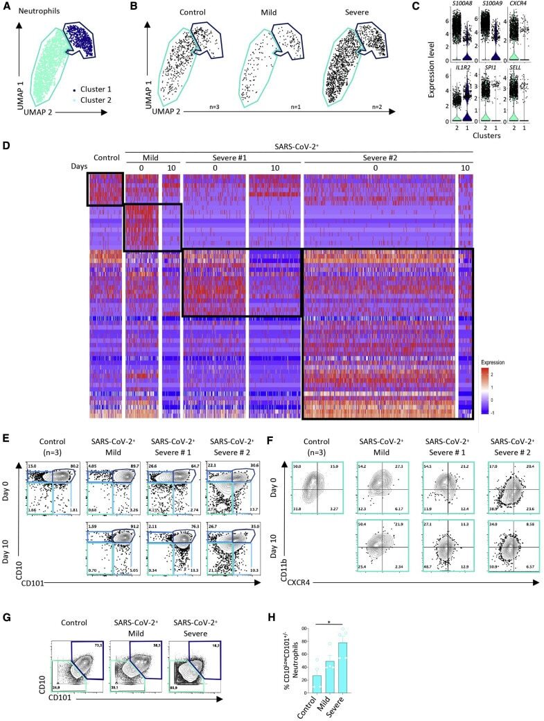
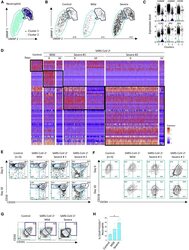
- Figure 4 Single-Cell Analysis of Neutrophils by scRNA-Seq, Spectral Flow Cytometry, and Mass Cytometry (A) UMAP profile of neutrophils in the 9 samples analyzed as described in Figure 2 A. (B) UMAP profile of neutrophils within the 3 controls and the mild and the two severe cases with the cluster gates overlaid. (C) Violin plots of expression of the indicated genes in two statistically defined neutrophil clusters. (D) Heatmap of DEGs in total neutrophils generated as described in Figure 3 B. (E and F) Spectral flow analysis of neutrophil subsets in pooled controls and each individual patient sample at day 0 and day 10, based on CD10 and CD101 expression (E) and CXCR4 and CD11b expression among CD10 Low CD101 - neutrophils (F) in the indicated samples (pooled controls). (G and H) Mass cytometry analysis of neutrophil subsets in 4 patients within each group (pooled data) as in Figures 3 F-3I, based on CD10 and CD101 expression (G) and the fraction of CD10 Low CD101 - neutrophils among total neutrophils in each sample within the 3 groups (H). Kruskal-Wallis test, * p < 0.05.