Explore
 Validate
 Learn
Learn Western blot
Antibody data
- Antibody Data
- Antigen structure
- References [2]
- Comments [0]
- Validations
- Western blot [1]
- Immunocytochemistry [2]
Submit
Validation data
Reference
Comment
Report error
- Product number
- 710428 - Provider product page

- Provider
- Invitrogen Antibodies
- Product name
- Cyclin D1 Recombinant Polyclonal Antibody (17HCLC)
- Antibody type
- Polyclonal
- Antigen
- Synthetic peptide
- Description
- Recombinant rabbit polyclonal antibodies are unique offerings from Thermo Fisher Scientific. They are comprised of a selection of multiple different recombinant monoclonal antibodies, providing the best of both worlds - the sensitivity of polyclonal antibodies with the specificity of monoclonal antibodies - all delivered with the consistency only found in a recombinant antibody. While functionally the same as a polyclonal antibody - recognizing multiple epitope sites on the target and producing higher detection sensitivity for low abundance targets - a recombinant rabbit polyclonal antibody has a known mixture of light and heavy chains. The exact population can be produced in every lot, circumventing the biological variability typically associated with polyclonal antibody production.
- Reactivity
- Human
- Host
- Rabbit
- Isotype
- IgG
- Antibody clone number
- 17HCLC
- Vial size
- 100 µg
- Concentration
- 0.5 mg/mL
- Storage
- Store at 4°C short term. For long term storage, store at -20°C, avoiding freeze/thaw cycles.
Submitted references Parthenolide inhibits the proliferation and induces the apoptosis of human uveal melanoma cells.
The molecular mechanism of treating osteoarthritis with dipsacus saponins by inhibiting chondrocyte apoptosis.
Che ST, Bie L, Li X, Qi H, Yu P, Zuo L
International journal of ophthalmology 2019;12(10):1531-1538
International journal of ophthalmology 2019;12(10):1531-1538
The molecular mechanism of treating osteoarthritis with dipsacus saponins by inhibiting chondrocyte apoptosis.
Li XR, Li J, Ren Q, Sun S
Experimental and therapeutic medicine 2017 Nov;14(5):4527-4532
Experimental and therapeutic medicine 2017 Nov;14(5):4527-4532
No comments: Submit comment
Supportive validation
- Submitted by
- Invitrogen Antibodies (provider)
- Main image

- Experimental details
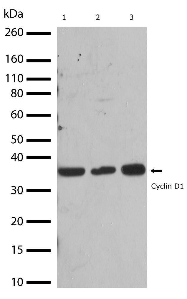
- Western blot analysis of Cyclin D1 in whole cell extracts from HeLa, A549, and MCF-7 (lanes 1-3 respectively) using a Cyclin D1 Recombinant Rabbit Polyclonal Antibody (Product # 710428) at a dilution of 2 µg/mL. Detection was performed using an HRP-conjugated Goat anti-Rabbit secondary antibody followed by chemiluminescence (ECL). Results show a band at ~36kDa.
Supportive validation
- Submitted by
- Invitrogen Antibodies (provider)
- Main image

- Experimental details
- Immunofluorescent analysis of Cyclin D1 in HeLa cells using a Cyclin D1 Recombinant Rabbit Polyclonal Antibody (Product # 710428) followed by detection using an Alexa Fluor 488-conjugated Goat anti-Rabbit secondary antibody (green) (Image A). Nuclei were stained using DAPI (Image B) and actin stained with Alexa Fluor 594 phalloidin (red) (image C). Image D is a composite image showing cytoplasmic and nuclear localization of Cyclin D1.

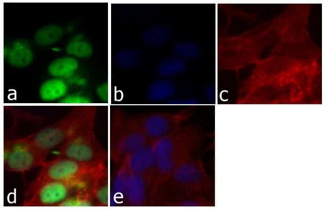
- Submitted by
- Invitrogen Antibodies (provider)
- Main image

- Experimental details
- Immunofluorescent analysis of Cyclin D1 was done on 70% confluent log phase HeLa cells. The cells were fixed with 4% paraformaldehyde for 15 minutes; permeabilized with 0.25% Triton X-100 for 10 minutes followed by blocking with 5% BSA for 1 hour at room temperature. The cells were incubated with Cyclin D1 Recombinant Rabbit Polyclonal Antibody (Product # 710428) at 1 µg\mL in 1% BSA and incubated for 3 hours at room temperature and then labeled with Alexa Fluor 488 Goat anti-Rabbit IgG Secondary Antibody (Product # A-11008) at a dilution of 1:400 for 30 minutes at room temperature (Panel a: green). Nuclei (Panel b: blue) were stained with SlowFade Gold Antifade Mountant with DAPI (Product # S36938). F-actin (Panel c: red) was stained with Alexa Fluor 594 Phalloidin (Product # A12381). Panel d is a merged image showing nuclear localization of Cyclin D1. Panel e shows no primary antibody. The images were captured at 20X magnification.