Explore
 Validate
 Learn
Learn Western blot
Antibody data
- Antibody Data
- Antigen structure
- References [3]
- Comments [0]
- Validations
- Western blot [1]
- Immunocytochemistry [1]
- Immunohistochemistry [3]
- Chromatin Immunoprecipitation [1]
Submit
Validation data
Reference
Comment
Report error
- Product number
- MA5-14585 - Provider product page

- Provider
- Invitrogen Antibodies
- Product name
- PAX5 Monoclonal Antibody (SP34)
- Antibody type
- Monoclonal
- Antigen
- Synthetic peptide
- Description
- MA5-14585 targets Pax-5 in IHC (P) applications and shows reactivity with Human and Mouse samples.
- Antibody clone number
- SP34
- Concentration
- 0.067 mg/mL
Submitted references A 2-Step Laemmli and Antigen Retrieval Method Improves Immunodetection.
Increased utilization, verification, and clinical implications of immunocytochemistry: Experience in a northern New England hospital.
Composite lymphoma in the anterior mediastinum: a case report and review of the literature.
Scalia CR, Gendusa R, Cattoretti G
Applied immunohistochemistry & molecular morphology : AIMM 2016 Jul;24(6):436-46
Applied immunohistochemistry & molecular morphology : AIMM 2016 Jul;24(6):436-46
Increased utilization, verification, and clinical implications of immunocytochemistry: Experience in a northern New England hospital.
Sauter JL, Ambaye AB, Mount SL
Diagnostic cytopathology 2015 Sep;43(9):688-95
Diagnostic cytopathology 2015 Sep;43(9):688-95
Composite lymphoma in the anterior mediastinum: a case report and review of the literature.
Yu G, Kong L, Qu G, Zhang Q, Wang W, Jiang L
Diagnostic pathology 2011 Jul 6;6:60
Diagnostic pathology 2011 Jul 6;6:60
No comments: Submit comment
Supportive validation
- Submitted by
- Invitrogen Antibodies (provider)
- Main image
- Experimental details
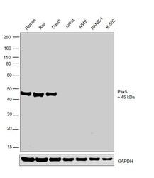
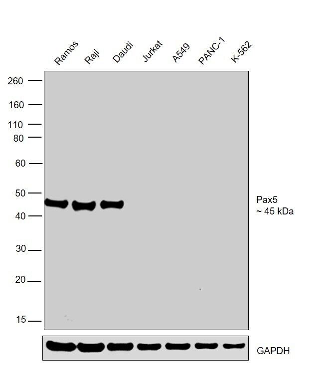
- Western blot was performed using Anti-PAX5 Monoclonal Antibody (SP34) (Product # MA5-14585) and a 45 kDa band corresponding to PAX5 was observed in Ramos, Raji and Daudi and not in Jurkat, A549, PANC-1 and K-562. Modified whole cell extracts (1% SDS) (30 µg lysate) of Ramos (Lane 1), Raji (Lane 2), Daudi (Lane 3), Jurkat (Lane 4), A549 (Lane 5), PANC-1 (Lane 6) and K-562 (Lane 7) were electrophoresed using Novex® NuPAGE® 4-12% Bis-Tris Protein Gel (Product # NP0322BOX). Resolved proteins were then transferred onto a nitrocellulose membrane (Product # IB23001) by iBlot® 2 Dry Blotting System (Product # IB21001). The blot was probed with the primary antibody (1:500 dilution) and detected by chemiluminescence with Goat anti-Rabbit IgG (H+L), Superclonal™ Recombinant Secondary Antibody, HRP (Product # A27036), using the iBright FL 1000 (Product # A32752). Chemiluminescent detection was performed using Novex® ECL Chemiluminescent Substrate Reagent Kit (Product # WP20005).
Supportive validation
- Submitted by
- Invitrogen Antibodies (provider)
- Main image

- Experimental details
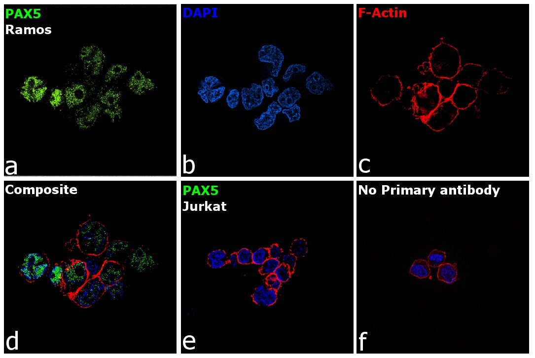
- Immunofluorescence analysis of PAX5 was performed using 70% confluent log phase Ramos cells. The cells were fixed with 4% Paraformaldehyde for 10 minutes, permeabilized with 0.1% Triton™ X-100 for 10 minutes, and blocked with 2% BSA for 10 minutes at room temperature. The cells were labeled with PAX5 Monoclonal Antibody (SP34) (Product # MA5-14585) at 1:250 dilution in 0.1% BSA, incubated at 4 degree Celsius overnight and then labeled with Goat anti-Rabbit IgG (H+L), Superclonal™ Recombinant Secondary Antibody, Alexa Fluor 488 (Product # A27034, 1:2000 dilution) for 45 minutes at room temperature (Panel a: Green). Nuclei (Panel b: Blue) were stained with SlowFade® Gold Antifade Mountant with DAPI (Product # S36938). F-actin (Panel c: Red) was stained with Rhodamine Phalloidin (Product # R415, 1:300). Panel d represents the merged image showing nuclear localization. Panel e represents Jurkat cells having no expression of PAX5. Panel f represents control cells with no primary antibody to assess background. The images were captured at 60X magnification.
Supportive validation
- Submitted by
- Invitrogen Antibodies (provider)
- Main image
- Experimental details
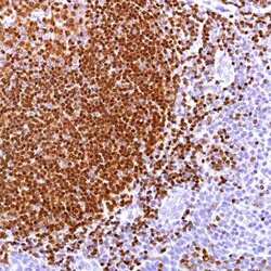
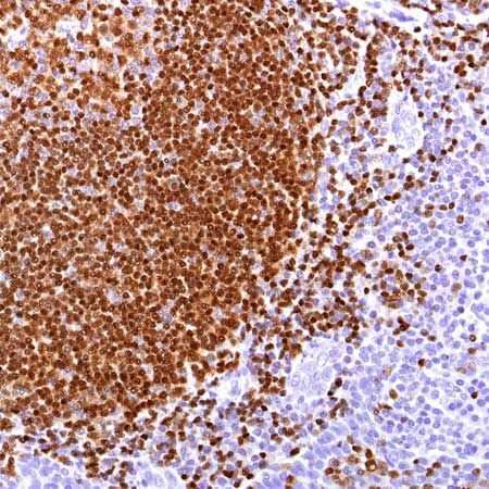
- Formalin-fixed, paraffin-embedded human Tonsil stained with PAX-5 using peroxidase-conjugate and DAB chromogen. Note nuclear staining.
- Submitted by
- Invitrogen Antibodies (provider)
- Main image
- Experimental details
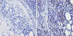
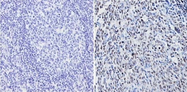
- Immunohistochemistry analysis of Pax-5 showing positive staining in the nucleus of paraffin-treated Human B lymphoma (right) compared with a negative control in the absence of primary antibody (left). To expose target proteins, antigen retrieval method was performed using 10mM sodium citrate (pH 6.0) microwaved for 8-15 min. Following antigen retrieval, tissues were blocked in 3% H2O2-methanol for 15 min at room temperature, washed with ddH2O and PBS, and then probed with a Pax-5 monoclonal antibody (Product # MA5-14585) diluted by 3% BSA-PBS at a dilution of 1:20 overnight at 4°C in a humidified chamber. Tissues were washed extensively PBST and detection was performed using an HRP-conjugated secondary antibody followed by colorimetric detection using a DAB kit. Tissues were counterstained with hematoxylin and dehydrated with ethanol and xylene to prep for mounting.
- Submitted by
- Invitrogen Antibodies (provider)
- Main image

- Experimental details
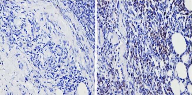
- Immunohistochemistry analysis of Pax-5 showing positive staining in the nucleus of paraffin-treated Human tonsil tissue (right) compared with a negative control in the absence of primary antibody (left). To expose target proteins, antigen retrieval method was performed using 10mM sodium citrate (pH 6.0) microwaved for 8-15 min. Following antigen retrieval, tissues were blocked in 3% H2O2-methanol for 15 min at room temperature, washed with ddH2O and PBS, and then probed with a Pax-5 monoclonal antibody (Product # MA5-14585) diluted by 3% BSA-PBS at a dilution of 1:20 overnight at 4°C in a humidified chamber. Tissues were washed extensively PBST and detection was performed using an HRP-conjugated secondary antibody followed by colorimetric detection using a DAB kit. Tissues were counterstained with hematoxylin and dehydrated with ethanol and xylene to prep for mounting.
Supportive validation
- Submitted by
- Invitrogen Antibodies (provider)
- Main image

- Experimental details
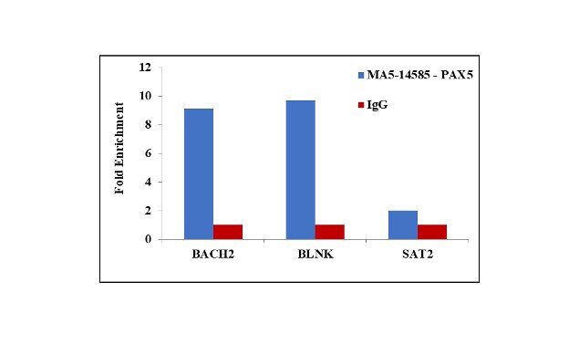
- Chromatin Immunoprecipitation (ChIP) assay of endogenous PAX5 protein using Anti-PAX5 Antibody: ChIP was performed using Anti-PAX5 Monoclonal Antibody (Product # MA5-14585, 5 µg) on sheared chromatin from Raji cells using the MAGnify ChIP System kit (Product # 49-2024). Normal Rabbit IgG was used as a negative IP control. The purified DNA was analyzed by qPCR using primers binding to BACH2 and BLNK promoter and SAT2 satellite repeats. Data is presented as fold enrichment of the antibody signal versus the negative control IgG using the comparative CT method.