Explore
 Validate
 Learn
Learn Western blot
 ELISA
ELISAAntibody data
- Antibody Data
- Antigen structure
- References [0]
- Comments [0]
- Validations
- Western blot [2]
Submit
Validation data
Reference
Comment
Report error
- Product number
- GTX48734 - Provider product page

- Provider
- GeneTex
- Proper citation
- GeneTex Cat#GTX48734, RRID:AB_11165825
- Product name
- Chk1 Yeast antibody
- Antibody type
- Polyclonal
- Reactivity
- Yeast
- Host
- Rabbit
- Storage
- Store vial at -20°C prior to opening. Aliquot contents and freeze at -20°C or below for extended storage. Avoid cycles of freezing and thawing.
No comments: Submit comment
Supportive validation
- Submitted by
- GeneTex (provider)
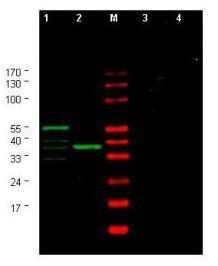
- Main image

- Experimental details
- Western blot using GeneTex's Affinity Purified anti-Yeast CHK1 antibody shows detection of a bands corresponding to CHK1 in Saccharomyces cerevisiae lysates. Two strains of S.cerevisiae were tested. Lane 1 shows a predominant band at ~60 kDa. Lane 2 shows a predominant band at ~38 kDa. Specific band staining is blocked when antibody is preincubated for 45 min at room temperature with 50 µg of peptide immunogen (lanes 3 and 4 respectively). Lysates were separated by 4-20% SDS-PAGE and transferred onto nitrocellulose. After blocking, the membrane was probed for 2 h at room temperature with the primary antibody diluted to 1:750 in blocking buffer diluted 1:5 in PBS. The membrane was washed and reacted with a 1:10,000 dilution of IRDye800 conjugated goat anti-Rabbit IgG [H&L] MX for 45 min at room temperature (800 nm channel, green). Molecular weight estimation was made by comparison to prestained MW markers in lane M (700 nm channel, red). IRDye800 fluorescence image was captured using the Odyssey® Infrared Imaging System developed by LI-COR. IRDye is a trademark of LI-COR, Inc. Other detection systems will yield similar results.
- Validation comment
- WB
- Submitted by
- GeneTex (provider)
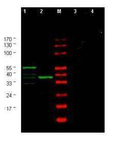
- Main image

- Experimental details
- Western blot using GeneTex's Affinity Purified anti-Yeast CHK1 antibody shows detection of a bands corresponding to CHK1 in Saccharomyces cerevisiae lysates. Two strains of S.cerevisiae were tested. Lane 1 shows a predominant band at ~60 kDa. Lane 2 shows a predominant band at ~38 kDa. Specific band staining is blocked when antibody is preincubated for 45 min at room temperature with 50 ?g of peptide immunogen (lanes 3 and 4 respectively). Lysates were separated by 4-20% SDS-PAGE and transferred onto nitrocellulose. After blocking, the membrane was probed for 2 h at room temperature with the primary antibody diluted to 1:750 in blocking buffer diluted 1:5 in PBS. The membrane was washed and reacted with a 1:10,000 dilution of IRDye800 conjugated goat anti-Rabbit IgG [H&L] MX for 45 min at room temperature (800 nm channel, green). Molecular weight estimation was made by comparison to prestained MW markers in lane M (700 nm channel, red). IRDye800 fluorescence image was captured using the Odyssey? Infrared Imaging System developed by LI-COR. IRDye is a trademark of LI-COR, Inc. Other detection systems will yield similar results.