 Explore
 Validate
 Learn
Learn Western blot
Antibody data
- Antibody Data
- Antigen structure
- References [2]
- Comments [0]
- Validations
- Western blot [1]
- Immunohistochemistry [3]
Submit
Validation data
Reference
Comment
Report error
- Product number
- AF4148 - Provider product page

- Provider
- Novus Biologicals
- Product name
- Goat Polyclonal HSPA8/HSC71/Hsc70 Antibody
- Antibody type
- Polyclonal
- Description
- Antigen Affinity-purified. Detects human, mouse and rat HSPA8/HSC71, Isoform 1 in Western blots. In Western blots, this antibody shows no cross-reactivity with recombinant human HSPA1A/HSP70, HSPA2, HSPA6, GRP75 or GRP78.
- Reactivity
- Human, Mouse, Rat
- Host
- Goat
- Conjugate
- Unconjugated
- Isotype
- IgG
- Vial size
- 100 ug
- Concentration
- LYOPH
- Storage
- Use a manual defrost freezer and avoid repeated freeze-thaw cycles. 12 months from date of receipt, -20 to -70 degreesC as supplied. 1 month, 2 to 8 degreesC under sterile conditions after reconstitution. 6 months, -20 to -70 degreesC under sterile conditions after reconstitution.
Submitted references Oncolytic Group B Adenovirus Enadenotucirev Mediates Non-apoptotic Cell Death with Membrane Disruption and Release of Inflammatory Mediators.
A sensitive and quantitative technique for detecting autophagic events based on lysosomal delivery.
Dyer A, Di Y, Calderon H, Illingworth S, Kueberuwa G, Tedcastle A, Jakeman P, Chia SL, Brown A, Silva MA, Barlow D, Beadle J, Hermiston T, Ferguson DJ, Champion B, Fisher KD, Seymour LW
Molecular therapy oncolytics 2017 Mar 17;4:18-30
Molecular therapy oncolytics 2017 Mar 17;4:18-30
A sensitive and quantitative technique for detecting autophagic events based on lysosomal delivery.
Katayama H, Kogure T, Mizushima N, Yoshimori T, Miyawaki A
Chemistry & biology 2011 Aug 26;18(8):1042-52
Chemistry & biology 2011 Aug 26;18(8):1042-52
No comments: Submit comment
Supportive validation
- Submitted by
- Novus Biologicals (provider)
- Main image
- Experimental details
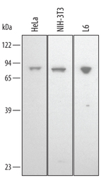
- Detection of Human/Mouse/Rat HSPA8/HSC71 by Western Blot. Western blot shows lysates of HeLa human cervical epithelial carcinoma cell line, NIH-3T3 mouse embryonic fibroblast cell line, and L6 rat myoblast cell line. PVDF membrane was probed with 1 µg/mL of Goat Anti-Human/Mouse/Rat HSPA8/HSC71 Antigen Affinity-purified Polyclonal Antibody (Catalog # AF4148) followed by HRP-conjugated Anti-Goat IgG Secondary Antibody (Catalog # HAF017). A specific band was detected for HSPA8/HSC71 at approximately 71 kDa (as indicated). This experiment was conducted using Immunoblot Buffer Group 2.

Supportive validation
- Submitted by
- Novus Biologicals (provider)
- Main image

- Experimental details
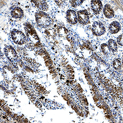
- HSPA8/HSC71 in Mouse Intestine. HSPA8/HSC71 was detected in immersion fixed frozen sections of mouse intestine using Goat Anti-Human/Mouse/Rat HSPA8/HSC71 Antigen Affinity-purified Polyclonal Antibody (Catalog # AF4148) at 3 µg/mL for 1 hour at room temperature followed by incubation with the Anti-Goat IgG VisUCyte™ HRP Polymer Antibody (Catalog # VC004). Tissue was stained using DAB (brown) and counterstained with hematoxylin (blue). Specific staining was localized to epithelial cells in intestinal glands. View our protocol for IHC Staining with VisUCyte HRP Polymer Detection Reagents.
- Submitted by
- Novus Biologicals (provider)
- Main image
- Experimental details
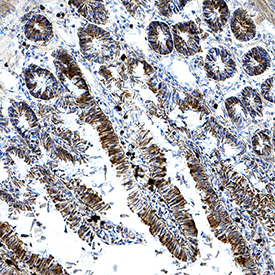
- HSPA8/HSC71 in Rat Intestine. HSPA8/HSC71 was detected in immersion fixed frozen sections of rat intestine using Goat Anti-Human/Mouse/Rat HSPA8/HSC71 Antigen Affinity-purified Polyclonal Antibody (Catalog # AF4148) at 3 µg/mL for 1 hour at room temperature followed by incubation with the Anti-Goat IgG VisUCyte™ HRP Polymer Antibody (Catalog # VC004). Tissue was stained using DAB (brown) and counterstained with hematoxylin (blue). Specific staining was localized to epithelial cells in intestinal glands. View our protocol for IHC Staining with VisUCyte HRP Polymer Detection Reagents.
- Submitted by
- Novus Biologicals (provider)
- Main image
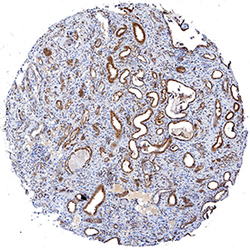
- Experimental details
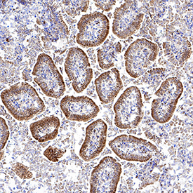
- HSPA8/HSC71 in Human Kidney. HSPA8/HSC71 was detected in immersion fixed paraffin-embedded sections of human kidney using Goat Anti-Human/Mouse/Rat HSPA8/HSC71 Antigen Affinity-purified Polyclonal Antibody (Catalog # AF4148) at 3 µg/mL for 1 hour at room temperature followed by incubation with the Anti-Goat IgG VisUCyte™ HRP Polymer Antibody (Catalog # VC004). Tissue was stained using DAB (brown) and counterstained with hematoxylin (blue). Specific staining was localized to epithelial cells in convoluted tubules. View our protocol for IHC Staining with VisUCyte HRP Polymer Detection Reagents.
